

Age alterations in extent and severity of experimental intranasal infection with Chlamydophila pneumoniae in BALB/c mice
- PMID: 15731073
- PMCID: PMC1064908
- DOI: 10.1128/IAI.73.3.1723-1734.2005
Age alterations in extent and severity of experimental intranasal infection with Chlamydophila pneumoniae in BALB/c mice
Abstract
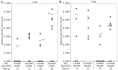
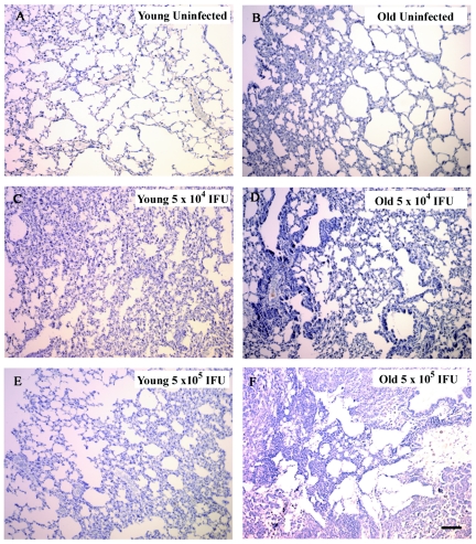
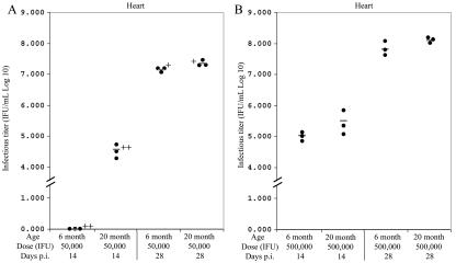
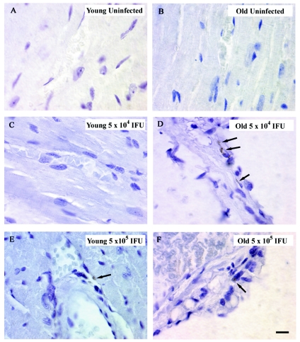
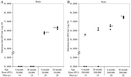
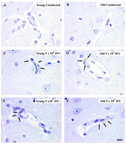
The intracellular bacterium Chlamydophila ("Chlamydia") pneumoniae is a pathogen for several respiratory diseases and may be a factor in the pathogenesis of chronic diseases of aging including atherosclerosis and Alzheimer's disease. We assessed whether aging is coupled with increased burden of infection in BALB/c mice after intranasal infection by C. pneumoniae. Six- and twenty-month-old BALB/c mice were infected intranasally with 5 x 10(4) inclusion forming units (IFU) or 5 x 10(5) IFU of C. pneumoniae. Lung, brain, and heart tissue were analyzed for infectious C. pneumoniae and for Chlamydophila antigen by immunohistochemistry. At both doses, aging was associated with a decreased proportion of animals that cleared infection from the lung and greater burden of infectious organism within the lung. We observed dose-dependent spread to the heart/ascending aorta in animals infected with C. pneumoniae. In mice given 5 x 10(4) IFU, spread to the heart by day 14 was only observed in old mice. By day 28, all animals inoculated with 5 x 10(4) IFU showed evidence of spread to the heart, although higher C. pneumoniae titers were observed in the hearts from old mice. In mice inoculated with 5 x 10(5) IFU, spread of C. pneumoniae to the heart was evident by day 14, with no discernible age effect. C. pneumoniae was also recovered from the central nervous system (brain and olfactory bulb) of all mice by day 28 postinfection, with higher C. pneumoniae titers in old animals than in young animals. Our results suggest that infection with C. pneumoniae may be more severe in old animals.
Figures

Similar articles
-
Severity of allergic airway disease due to house dust mite allergen is not increased after clinical recovery of lung infection with Chlamydia pneumoniae in mice.Infect Immun. 2013 Sep;81(9):3366-74. doi: 10.1128/IAI.00334-13. Epub 2013 Jul 1. Infect Immun. 2013. PMID: 23817611 Free PMC article.
-
Chlamydia pneumoniae infection does not induce or modify atherosclerosis in mice.Circulation. 2001 Jun 12;103(23):2834-8. doi: 10.1161/01.cir.103.23.2834. Circulation. 2001. PMID: 11401941
-
Detection of Chlamydophila pneumoniae in mouse respiratory ciliated epithelium using targeted sections of the lung tissue.Bull Exp Biol Med. 2002 Nov;134(5):460-2. doi: 10.1023/a:1022642414632. Bull Exp Biol Med. 2002. PMID: 12802452
-
Chlamydia pneumoniae and atherosclerosis.Semin Respir Infect. 2003 Mar;18(1):48-54. doi: 10.1053/srin.2003.50006. Semin Respir Infect. 2003. PMID: 12652454 Review.
-
Chlamydia pneumoniae as a respiratory pathogen.Front Biosci. 2002 Mar 1;7:e66-76. doi: 10.2741/hahn. Front Biosci. 2002. PMID: 11861211 Review.
Cited by
-
Detection of bacterial antigens and Alzheimer's disease-like pathology in the central nervous system of BALB/c mice following intranasal infection with a laboratory isolate of Chlamydia pneumoniae.Front Aging Neurosci. 2014 Dec 5;6:304. doi: 10.3389/fnagi.2014.00304. eCollection 2014. Front Aging Neurosci. 2014. PMID: 25538615 Free PMC article.
-
Chlamydia pneumoniae infection in atherosclerotic lesion development through oxidative stress: a brief overview.Int J Mol Sci. 2013 Jul 19;14(7):15105-20. doi: 10.3390/ijms140715105. Int J Mol Sci. 2013. PMID: 23877837 Free PMC article. Review.
-
Chlamydia pneumoniae infection and Alzheimer's disease: a connection to remember?Med Microbiol Immunol. 2010 Nov;199(4):283-9. doi: 10.1007/s00430-010-0162-1. Epub 2010 May 6. Med Microbiol Immunol. 2010. PMID: 20445987 Review.
-
Chlamydia pneumoniae Clinical Isolate from Gingival Crevicular Fluid: A Potential Atherogenic Strain.Front Cell Infect Microbiol. 2015 Nov 25;5:86. doi: 10.3389/fcimb.2015.00086. eCollection 2015. Front Cell Infect Microbiol. 2015. PMID: 26636048 Free PMC article.
-
Chlamydia pneumoniae in Alzheimer's disease pathology.Front Neurosci. 2024 May 6;18:1393293. doi: 10.3389/fnins.2024.1393293. eCollection 2024. Front Neurosci. 2024. PMID: 38770241 Free PMC article. Review.
References
-
- Abrams, J. T., B. J. Balin, and E. C. Vonderheid. 2001. Association between Sezary T cell-activating factor, Chlamydia pneumoniae, and cutaneous T-cell lymphoma. Ann. N. Y. Acad. Sci. 941:69-85. - PubMed
-
- Airenne, S., H. M. Surcel, A. Bloigu, K. Laitinen, P. Saikku, and A. Laurila. 2000. The resistance of human monocyte-derived macrophages to Chlamydia pneumoniae infection is enhanced by interferon-gamma. Apmis 108:139-144. - PubMed
-
- Anton, E., A. Otegui, and A. Alonso. 2000. Meningoencephalitis and Chlamydia pneumoniae infection. Eur. J. Neurol. 7:586. - PubMed
-
- Balin, B. J., H. C. Gerard, E. J. Arking, D. M. Appelt, P. J. Branigan, J. T. Abrams, J. A. Whittum-Hudson, and A. P. Hudson. 1998. Identification and localization of Chlamydia pneumoniae in the Alzheimer's brain. Med. Microbiol. Immunol. 187:23-42. - PubMed
Publication types
MeSH terms
Grants and funding
LinkOut - more resources
Full Text Sources
Other Literature Sources
Medical
