

B1 lymphocytes and myeloid dendritic cells in lymphoid organs are preferential extratumoral sites of parvovirus minute virus of mice prototype strain expression
- PMID: 15731246
- PMCID: PMC1075710
- DOI: 10.1128/JVI.79.6.3517-3524.2005
B1 lymphocytes and myeloid dendritic cells in lymphoid organs are preferential extratumoral sites of parvovirus minute virus of mice prototype strain expression
Abstract
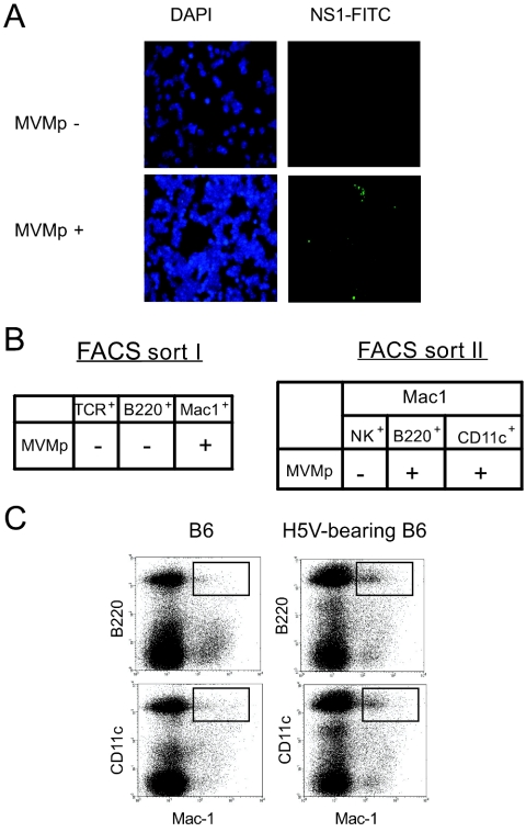
Due to their oncolytic properties and apathogenicity, autonomous parvoviruses have attracted significant interest as possible anticancer agents. Recent preclinical studies provided evidence of the therapeutic potential of minute virus of mice prototype strain (MVMp) and its recombinant derivatives. In a murine model of hemangiosarcoma, positive therapeutic outcome correlated with high intratumoral expression of MVMp-encoded genes in tumors and lymphoid organs, especially in tumor-draining lymph nodes. The source and relevance of this extratumoral expression, which came as a surprise because of the known fibrotropism of MVMp, remained unclear. In the present study, we investigated (i) whether the observed expression pattern occurs in different tumor models, (ii) which cell population is targeted by the virus, and (iii) the immunological consequences of this infection. Significant MVMp gene expression was detected in lymphoid tissues from infected tumor-free as well as melanoma-, lymphoma-, and hemangiosarcoma-bearing mice. This expression was especially marked in lymph nodes draining virus-injected tumors. Fluorescent in situ hybridization analysis, multicolor fluorescence-activated cell sorting, and quantitative reverse transcription-PCR revealed that MVMp was expressed in rare subpopulations of CD11b (Mac1)-positive cells displaying CD11c+ (myeloid dendritic cells [MDC]) or CD45B (B220+ [B1 lymphocytes]) markers. Apart from the late deletion of cytotoxic memory cells (CD8+ CD44+ CD62L-), this infection did not lead to significant alteration of the immunological profile of cells populating lymphoid organs. However, subtle changes were detected in the production of specific proinflammatory cytokines in lymph nodes from virus-treated animals. Considering the role of B1 lymphocytes and MDC in cancer and immunological surveillance, the specific ability of these cell types to sustain parvovirus-driven gene expression may be exploited in gene therapy protocols.
Figures


Similar articles
-
Humoral immune responses against minute virus of mice vectors.J Gene Med. 2006 Sep;8(9):1141-50. doi: 10.1002/jgm.940. J Gene Med. 2006. PMID: 16800041
-
Myeloid marker expression on antiviral CD8+ T cells following an acute virus infection.Eur J Immunol. 2003 Oct;33(10):2736-43. doi: 10.1002/eji.200324087. Eur J Immunol. 2003. PMID: 14515257
-
Anatomic location defines antigen presentation by dendritic cells to T cells in response to intravenous soluble antigens.Eur J Immunol. 2007 Jun;37(6):1453-62. doi: 10.1002/eji.200636544. Eur J Immunol. 2007. PMID: 17474148
-
Artificial engineering of secondary lymphoid organs.Adv Immunol. 2010;105:131-57. doi: 10.1016/S0065-2776(10)05005-4. Adv Immunol. 2010. PMID: 20510732 Review.
-
Organogenesis of lymphoid tissues.Nat Rev Immunol. 2003 Apr;3(4):292-303. doi: 10.1038/nri1054. Nat Rev Immunol. 2003. PMID: 12669020 Review.
Cited by
-
Toll-like receptor 9 in plasmacytoid dendritic cells fails to detect parvoviruses.J Virol. 2013 Mar;87(6):3605-8. doi: 10.1128/JVI.03155-12. Epub 2013 Jan 9. J Virol. 2013. PMID: 23302877 Free PMC article.
-
Immune System Stimulation by Oncolytic Rodent Protoparvoviruses.Viruses. 2019 May 4;11(5):415. doi: 10.3390/v11050415. Viruses. 2019. PMID: 31060205 Free PMC article. Review.
-
Immunological mediators in the causal relationship between latent autoimmune diabetes in adults and breast cancer: A Mendelian randomization analysis.Medicine (Baltimore). 2025 May 23;104(21):e42571. doi: 10.1097/MD.0000000000042571. Medicine (Baltimore). 2025. PMID: 40419887 Free PMC article.
-
Parvoviruses-tools to fine-tune anticancer immune responses.Oncoimmunology. 2012 Nov 1;1(8):1417-1419. doi: 10.4161/onci.21097. Oncoimmunology. 2012. PMID: 23243613 Free PMC article.
-
Distinct host cell fates for human malignant melanoma targeted by oncolytic rodent parvoviruses.Virology. 2013 Nov;446(1-2):37-48. doi: 10.1016/j.virol.2013.07.013. Epub 2013 Aug 9. Virology. 2013. PMID: 24074565 Free PMC article.
References
-
- Angiolillo, A. L., C. Sgadari, and G. Tosato. 1996. A role for the interferon-inducible protein 10 in inhibition of angiogenesis by interleukin-12. Ann. N. Y. Acad. Sci. 795:158-167. - PubMed
-
- Barker, P. E., L. Savelyeva, and M. Schwab. 1993. Translocation junctions cluster at the distal short arm of chromosome 1 (1p36.1-2) in human neuroblastoma cells. Oncogene 8:3353-3358. - PubMed
-
- Bottazzi, B., S. Walter, D. Govoni, F. Colotta, and A. Mantovani. 1992. Monocyte chemotactic cytokine gene transfer modulates macrophage infiltration, growth, and susceptibility to IL-2 therapy of a murine melanoma. J. Immunol. 148:1280-1285. - PubMed
-
- Brownstein, D. G., A. L. Smith, E. A. Johnson, D. J. Pintel, L. K. Naeger, and P. Tattersall. 1992. The pathogenesis of infection with minute virus of mice depends on expression of the small nonstructural protein NS2 and on the genotype of the allotropic determinants VP1 and VP2. J. Virol. 66:3118-3124. - PMC - PubMed
Publication types
MeSH terms
Substances
LinkOut - more resources
Full Text Sources
Molecular Biology Databases
Research Materials
Miscellaneous
