. 2005 Mar;79(6):3615-26.
doi: 10.1128/JVI.79.6.3615-3626.2005.

Analysis of human cytomegalovirus oriLyt sequence requirements in the context of the viral genome

Affiliations

Analysis of human cytomegalovirus oriLyt sequence requirements in the context of the viral genome

Eva-Maria Borst et al. J Virol. 2005 Mar.

Abstract

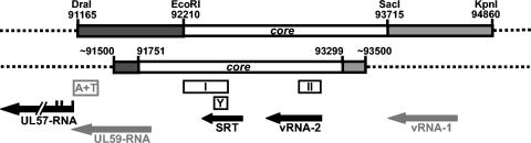
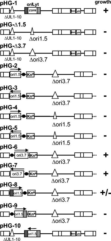
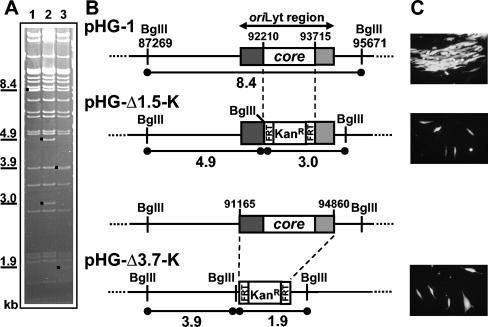
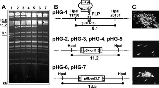
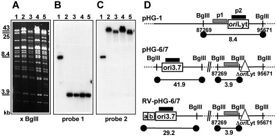
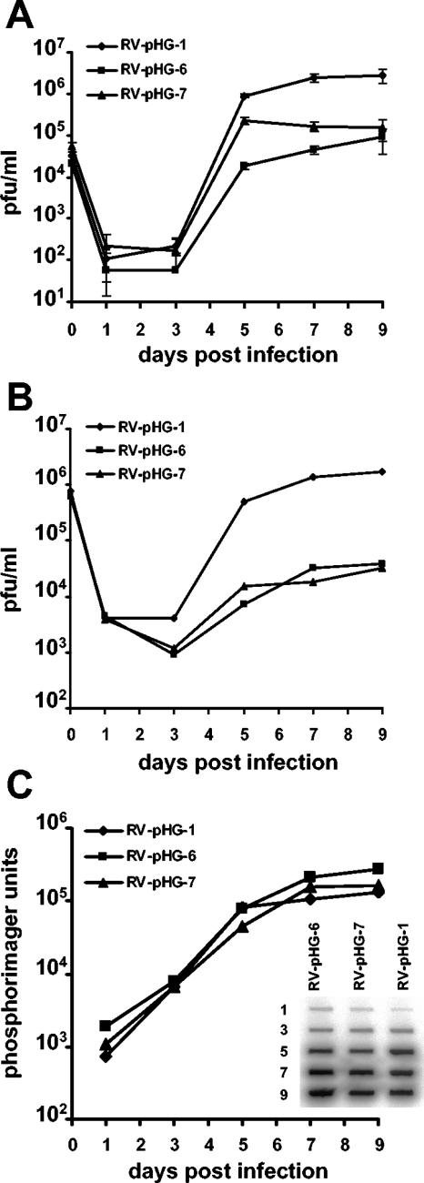
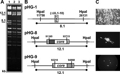
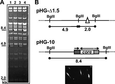
During the lytic phase of infection, replication of herpesvirus genomes initiates at the lytic origin of replication, oriLyt. Many herpesviruses harbor more than one lytic origin, but so far, only one oriLyt has been identified for human cytomegalovirus (HCMV). Evidence for the existence of additional lytic origins of HCMV has remained elusive. On the basis of transient replication assays with cloned viral fragments, HCMV oriLyt was described as a core region of 1.5 kbp (minimal oriLyt) flanked by auxiliary sequences required for maximal replication activity (complete oriLyt). It remained unclear whether minimal oriLyt alone can drive the replication of HCMV in the absence of its accessory regions. To investigate the sequence requirements of oriLyt in the context of the viral genome, mutant genomes were constructed lacking either minimal or complete oriLyt. These genomes were not infectious, suggesting that HCMV contains only one lytic origin of replication. Either minimal or complete oriLyt was then ectopically reinserted into the oriLyt-depleted genomes. Only the mutant genomes carrying complete oriLyt led to infectious progeny. Remarkably, inversion of the 1.5-kbp core origin relative to its flanking regions resulted in a replication-defective genome. Mutant genomes carrying minimal oriLyt plus the left flanking region gave rise to minifoci, but genomes harboring minimal oriLyt together with the right flanking region were noninfectious. We conclude that the previously defined minimal lytic origin is not sufficient to drive replication of the HCMV genome. Rather, our results underline the importance of the accessory regions and their correct arrangement for the function of HCMV oriLyt.


Figures

FIG. 1.
FIG. 1.
Schematic of the HCMV oriLyt region. Organization of oriLyt as determined by Masse et al. (upper line) and Zhu et al. (lower line). The origin core is depicted in white, and the left and right auxiliary regions are shown in dark and light grey, respectively. Nucleotide positions are according to Chee et al. Essential elements are shown in black, and elements supposed to be nonessential are in grey. A+T, A/T-rich region; I and II, essential regions I and II; Y, oligopyrimidine sequence. The arrows represent the transcripts detected within and around oriLyt. The 5′ start sites of the UL57 RNA are indicated. Not shown is the 6.5-kb RNA that crosses the origin region. Mutant genomes with a deletion of minimal oriLyt lack the origin core as defined by Masse et al. (nt 92210 to 93715), and mutant genomes with the complete lytic origin deleted lack the sequences between nt 91165 and 94860.
FIG. 2.
FIG. 2.
Schematic overview of recombinant HCMV genomes generated and tested in this study. Shown are the parental BAC pHG-1 (top line) and the mutant BACs harboring a deletion of either the minimal (Δori1.5) or the complete (Δori3.7) lytic origin. Ectopic insertion of minimal oriLyt (ori1.5), complete oriLyt (ori3.7), minimal oriLyt plus the left flanking region (dark grey), or minimal oriLyt plus the right flanking region (light grey) is indicated by rectangles on the left-hand side of the genomes. The arrows reflect the respective orientations of the origin sequences. Also shown are the locations of the backbone (0.5 kbp; black dots) and the kanamycin resistance marker (1.0 kbp; KnR) of the shuttle vector. pHG-10 represents a genome carrying an inversion of the origin core at its original position. The genomes are not drawn to scale. The growth properties of the respective genomes are illustrated to the right: −, no plaque formation after transfection, +, plaque formation leading to complete CPE; +/−, formation of minifoci.
FIG. 3.
FIG. 3.
Construction and analysis of HCMV genomes with deletion of the minimal or complete lytic origin. (A) Structural analysis of the parental BAC pHG-1 (lane 1) and mutant BACs pHG-Δ1.5K (lane 2) and pHG-Δ3.7K (lane 3) by treatment of the BAC DNA with BglII, followed by agarose gel electrophoresis and ethidium bromide staining. Relevant fragments are marked by black dots. Note that the 8.4-kbp band in lane 1 and the 3.9-kbp band in lane 3 are both comigrating with bands of similar size. (B) Schematic drawing of the genomes depicting the deletion of either minimal (core; pHG-Δ1.5-K) or complete (pHG-Δ3.7-K) oriLyt. The sizes of fragments characteristic of the different BACs are indicated below each diagram. Dark grey, accessory region flanking the origin core to the left; light grey, accessory region flanking the core origin to the right; KanR, kanamycin resistance marker. (C) Human fibroblasts transfected with pHG-1, pHG-Δ1.5, or pHG-Δ3.7 analyzed 10 days posttransfection by UV light microscopy.
FIG. 4.
FIG. 4.
Ectopic reinsertion of minimal or complete oriLyt. (A) Restriction analysis with HpaI of pHG-1 (lane 1), recombinant BACs pHG-4 and pHG-5 (lanes 2 and 3) derived from pHG-Δ1.5 and harboring minimal oriLyt in a different orientation at the ectopic position, and pHG-2, pHG-3 (lanes 4 and 5), and pHG-6 and pHG-7 (lanes 6 and 7) constructed by ectopic insertion of minimal oriLyt and complete oriLyt into pHG-Δ3.7 in different orientations. Relevant DNA bands are indicated. (B) Structure of the ectopic region before (pHG-1) and after insertion of the respective origin sequences (pHG-2 to pHG-7). Also depicted is the FLP-mediated integration of the shuttle plasmids into the FRT site at the ectopic position (top diagram). (C) Analysis of plaque formation on human fibroblasts transfected with either the parental or the respective mutant genomes. The micrographs show cell cultures at 10 days posttransfection with pHG-1 (top), pHG-2 (middle), and pHG-6 (bottom). Results virtually identical to those for pHG-2 and pHG-6 were obtained after transfection of pHG-3, pHG-4, or pHG-5, and of pHG-7, respectively.
FIG. 5.
FIG. 5.
Structural analysis of BACs pHG-6 and pHG-7 and of the genomes of the corresponding viruses RV-pHG-6 and RV-pHG-7. (A) Characterization of BAC DNAs (lane 1, pHG-1; lane 2, pHG-6; lane 4, pHG-7) and of viral DNAs (lane 3, RV-pHG-6; lane 5, RV-pHG-7) by BglII restriction analysis and agarose gel electrophoresis. The sizes of relevant DNA fragments are indicated. (B and C) Southern blot analysis with probes p1 and p2. (D) Schematic drawing of the predicted genomic regions representing the locations of oriLyt in the parental (top scheme; pHG-1) and mutant (second scheme; pHG-6 and pHG-7) BACs and in the corresponding virus genomes (bottom scheme; RV-pHG-6 and RV-pHG-7). The locations of the probes used for hybridization are shown by black and grey bars, and the sizes of the hybridizing fragments are depicted below each diagram. The 29.2-kbp terminal fragment depicted in the bottom scheme arises from linear virus genomes with the UL region in the prototype orientation and represents one of three different fragments detected by probe p2 (see the text).
FIG. 6.
FIG. 6.
Growth analysis of the parental virus RV-pHG-1 and of the mutant viruses RV-pHG-6 and RV-pHG-7. (A and B) HFF were infected at an MOI of 0.025 (A) or 1.5 (B), and at the indicated times postinfection, supernatants of the infected cultures were analyzed for infectious virus by plaque assay. Each data point represents the average of three independent wells. Titers at day zero represent input inocula. (C) Amplification of viral DNA after infection. HFF were infected at an MOI of 0.02 PFU/cell, and total DNA was prepared on days 1, 3, 5, 7, and 9 postinfection. The DNAs were assayed by slot blotting using an HCMV-specific probe, followed by quantification of the radioactivity using a phosphorimager.
FIG. 7.
FIG. 7.
Ectopic insertion of the origin core together with either its left or right flanking region. (A) DNAs of pHG-1, pHG-8, and pHG-9 (lanes 1 to 3) were cut with HpaI and separated on a 0.5% agarose gel, followed by ethidium bromide staining. (B) Structure of pHG-8 (middle line) and pHG-9 (bottom line) after FLP-mediated insertion of the indicated origin sequences. (C) HFF cells transfected with pHG-1, pHG-8, or pHG-9 were analyzed for plaque formation 3 weeks after transfection.
FIG. 8.
FIG. 8.
Inversion of the origin core at its original position in the HCMV genome. (A) Following mutagenesis, BAC DNA was isolated from several clones and checked for the presence of fragments indicative of the mutant genomes by restriction analysis with BglII. Lanes 1 and 3, DNAs of BAC clones with a structure identical to that of parental pHG-Δ1.5. Lanes 2 and 4, DNAs of BACs that acquired the origin core in inverted orientation. DNA bands characteristic of the different BACs are marked by black dots. (B) Structure of the HCMV oriLyt regions of pHG-Δ1.5 harboring a deletion of the origin core and of pHG-10 carrying the origin core in inverted orientation. Shown below are human fibroblasts transfected with pHG-10 3 weeks posttransfection.

Similar articles

Cited by

References

    1. Anders, D. G., M. A. Kacica, G. Pari, and S. M. Punturieri. 1992. Boundaries and structure of human cytomegalovirus oriLyt, a complex origin for lytic-phase DNA replication. J. Virol. 66:3373-3384. - PMC - PubMed
    1. Anders, D. G., and S. M. Punturieri. 1991. Multicomponent origin of cytomegalovirus lytic-phase DNA replication. J. Virol. 65:931-937. - PMC - PubMed
    1. Atalay, R., A. Zimmermann, M. Wagner, E. Borst, C. Benz, M. Messerle, and H. Hengel. 2002. Identification and expression of human cytomegalovirus transcription units coding for two distinct Fcγ receptor homologs. J. Virol. 76:8596-8608. - PMC - PubMed
    1. AuCoin, D. P., K. S. Colletti, Y. Xu, S. A. Cei, and G. S. Pari. 2002. Kaposi's sarcoma-associated herpesvirus (human herpesvirus 8) contains two functional lytic origins of DNA replication. J. Virol. 76:7890-7896. - PMC - PubMed
    1. Baker, A., and M. Cotten. 1997. Delivery of bacterial artificial chromosomes into mammalian cells with psoralen-inactivated adenovirus carrier. Nucleic Acids Res. 25:1950-1956. - PMC - PubMed

Publication types

LinkOut - more resources