. 2005 Mar;79(6):3703-12.
doi: 10.1128/JVI.79.6.3703-3712.2005.

BFRF1 of Epstein-Barr virus is essential for efficient primary viral envelopment and egress

Affiliations

BFRF1 of Epstein-Barr virus is essential for efficient primary viral envelopment and egress

Antonella Farina et al. J Virol. 2005 Mar.

Abstract

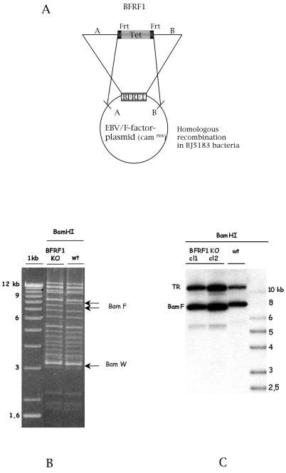
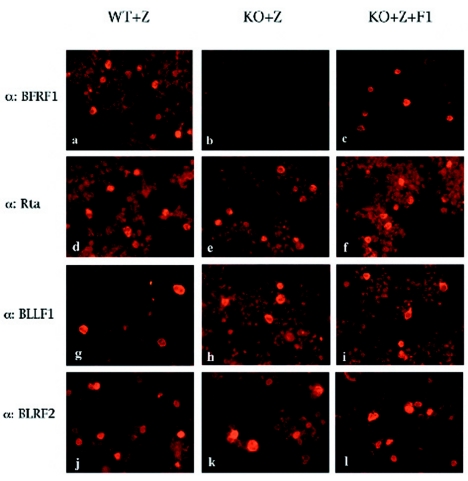
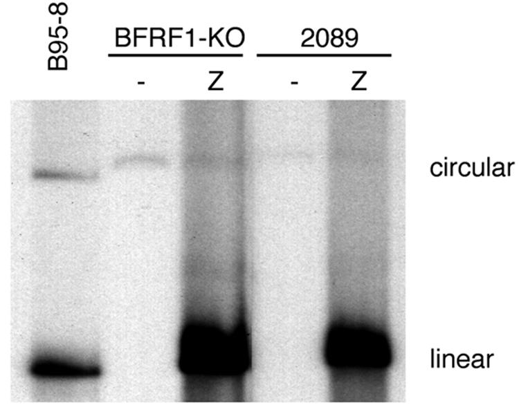
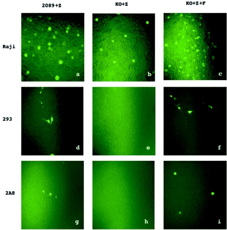
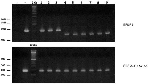
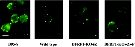
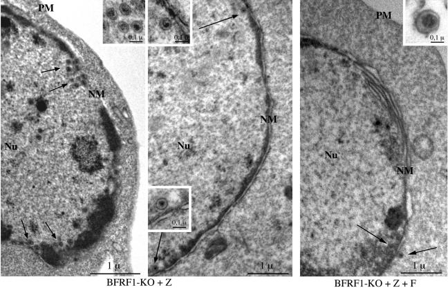
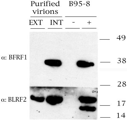
The molecular mechanisms that underlie maturation and egress of Epstein-Barr virus (EBV) virions are only partially characterized. We have recently shown that the BFRF1 gene, the EBV positional homolog of herpes simplex virus type 1 and pseudorabies virus UL34, is expressed early during EBV lytic replication and that it is found predominantly on the nuclear membrane (A. Farina, R. Santarelli, R. Gonnella, R. Bei, R. Muraro, G. Cardinali, S. Uccini, G. Ragona, L. Frati, A. Faggioni, and A. Angeloni, J. Virol. 74:3235-3244, 2000). These data suggest that the BFRF1 protein might be involved in viral primary envelopment. To precisely determine the function of this protein, we have constructed an EBV mutant devoid of the BFRF1 gene (BFRF1-KO). 293 cells carrying BFRF1-KO showed no differences in comparison with wild-type EBV in terms of DNA lytic replication or expression of late viral proteins upon induction of the lytic cycle. However, binding assays and infection experiments using cell lines or human cord blood lymphocytes showed a clear reduction in the viral mutant titers. Complementation experiments with BFRF1-KO and a BFRF1 expression vector restored viral titers to levels similar to those for the wild-type control, showing that the modifications that we introduced were limited to the BFRF1 gene. Electron microscopic observations showed that the reduction in viral titers was due to sequestration of EBV nucleocapsids in the nuclei of lytically induced cells. This suggests that BFRF1 is involved in transport of the maturing virion across the nuclear membrane. This hypothesis was further supported by the observation that BFRF1 is present in maturing intracellular virions but not in their extracellular counterparts. This implies that BFRF1 is a key protein for EBV maturation.


Figures

FIG. 1.
FIG. 1.
Mutant EBV lacking BFRF1. (A) Construction of the BFRF1-negative EBV mutant. Homologous recombination between the wild-type EBV genome and a linearized fragment carrying the BamHI F region in which 634 bp of the BFRF1 gene was replaced by the gene for the tetracycline resistance was performed in the recBCD E. coli strain BJ5183. Clones carrying the recombinant EBV were selected for chloramphenicol and tetracycline resistance. In order to excise the Tet box, selected clones were transformed with a plasmid encoding the Flp recombinase (pCP20) that recombined with the two Flp recombination target (Frt) sequences flanking the Tet box. (B) Restriction fragment analysis of EBV BFRF1-KO DNA. The restriction pattern of BFRF1-KO mutant DNA was compared to that of the wild-type (wt) EBV DNA. The modified DNA pattern is indicated by arrows. As expected from the predicted BamHI restriction pattern, the deletion of the BFRF1 fragment led to a shift of the BamHI F region with respect to that in the wild-type DNA. (C) Southern blot analysis of 293-BFRF1-KO cell clones compared to 293-2089 cells carrying the wild-type EBV genome. Genomic DNA from stably transfected clones harboring the BFRF1-KO mutant was extracted and cleaved with BamHI restriction enzyme. After agarose gel electrophoresis, Southern blot analysis was performed with a probe specific for the BFRF1 region and for the TR region. As expected, the BamHI F fragment showed a shift due to the deletion in the BFRF1 gene. In contrast, the TR fragment was unchanged, confirming that passaging in E. coli did not reduce the number of repeats within the virus.
FIG. 2.
FIG. 2.
Expression of early and late proteins in lytically induced 293-BFRF1-KO cell lines. The results of an immunofluorescence assay performed on lytically induced 293-2089, 293-BFRF1-KO, and BFRF1-complemented 293-BFRF1-KO cell lines are shown. Lytically induced 293-BFRF1-KO cells do not show any expression of BFRF1 (b) compared to the wild-type 293-2089 cells or to the BFRF1-complemented 293BFRF1-KO cells (a and c, respectively), while the expression of the immediate-early antigen Rta (e), as well as of the late antigens BLLF1 (h) and BLRF2 (k), is still maintained in the 293-BFRF1-KO cell line at a level comparable to that in the wild-type or the BFRF1 complemented 293-BFRF1-KO cells.
FIG. 3.
FIG. 3.
Gardella gel analysis of unit-length EBV DNA in 293-BFRF1-KO cells. Linear forms of the virus are detected in 293-BFRF1-KO cells upon induction of the lytic cycle as well as in 293-2089 producing cells that were used as a positive control. This experiment shows that BFRF1 deletion does not affect lytic viral DNA replication.
FIG. 4.
FIG. 4.
Infection of different cell types with supernatants from induced 293 cells containing wild-type EBV (2089+Z), BFRF1-KO (KO+Z), or complemented BFRF1-KO (KO+Z+F), as detected by GFP expression with an inverted epifluorescence microscope.
FIG. 5.
FIG. 5.
(Upper panel) PCR analysis for the BFRF1 gene of DNA from immortalized B-cell clones that were established after infection with wild-type EBV (lanes 1 to 3), with complemented BFRF1-KO EBV (lanes 4 to 6), or with BFRF1-KO EBV (lanes 7 to 9) and of control DNA from B95-8 cell lines (+) or from DG75 cell lines (−). (Lower panel) Control PCR analysis of the EBER-1 region.
FIG. 6.
FIG. 6.
Virus binding on the surface of Raji cells as detected by indirect immunofluorescence staining of the major envelope glycoprotein gp350/220. (a) EBV from supernatant of B95-8 cells; (b) supernatant of induced 293-2089 cells; (c) supernatant of 293-BFRF1-KO cells; (d) supernatant of BFRF1-complemented 293-BFRF1-KO cells. Bar, 10 μm.
FIG. 7.
FIG. 7.
Ultrastructural observations of induced 293-BFRF1-KO cells and 293-BFRF1-KO cells complemented with BFRF1. Left and center micrographs: numerous nucleocapsids are present in the nuclei of induced BFRF1-KO cells and are distributed mainly in the proximity of the nuclear membrane (arrows). Insets show an higher magnification of fully assembled intranuclear nucleocapsids containing electron-dense material corresponding to viral DNA. Right micrograph: few nucleocapsids (arrows) are visible in BFRF1-complemented cells. A typical multilayered reduplication of the nuclear membrane (NM) is shown. An extracellular mature virion is shown at higher magnification in the inset. PM, plasma membrane; Nu, nucleus. Bars, 1 μm (0.1 μm in the insets).
FIG. 8.
FIG. 8.
Western immunoblot analysis performed on purified virions. Fractions containing extracellular or intracellular purified virions were probed with antibodies directed against BFRF1 and BLRF2 gene products. An immunoblot of uninduced (−) and TPA-induced (+) B95-8 cell extracts is shown as a control.

References

    1. Angeloni, A., A. Farina, G. Gentile, A. Capobianchi, P. Martino, V. Visco, R. Muraro, L. Frati, and A. Faggioni. 2001. Epstein-Barr virus and breast cancer: search for antibodies to the novel BFRF1 protein in sera of breast cancer patients. J. Natl. Cancer Inst. 93:560-561. - PubMed
    1. Ben-Bassat, H., N. Goldblum, S. Mitrani, T. Goldblum, J. M. Yoffey, M. M. Cohen, Z. Bentwich, B. Ramot, E. Klein, and G. Klein. 1977. Establishment in continuous culture of a new type of lymphocyte from a “Burkitt like” malignant lymphoma (line D.G.-75). Int. J. Cancer 19:27-33. - PubMed
    1. Boukamp, P., R. T. Petrussevska, D. Breitkreutz, J. Hornung, A. Markham, and N. E. Fusenig. 1988. Normal keratinization in a spontaneously immortalized aneuploid human keratinocyte cell line. J. Cell Biol. 106:761-771. - PMC - PubMed
    1. Cherepanov, P. P., and W. Wackernagel. 1995. Gene disruption in Escherichia coli: TcR and KmR cassettes with the option of Flp-catalyzed excision of the antibiotic-resistance determinant. Gene 158:9-14. - PubMed
    1. Cho, M. S., G. Milman, and S. D. Hayward. 1985. A second Epstein-Barr virus early antigen gene in BamHI fragment M encodes a 48- to 50-kilodalton nuclear protein. J. Virol. 56:860-866. - PMC - PubMed

Publication types

MeSH terms

LinkOut - more resources