. 2005 Mar;79(6):3766-74.
doi: 10.1128/JVI.79.6.3766-3774.2005.

Importance of both the coding and the segment-specific noncoding regions of the influenza A virus NS segment for its efficient incorporation into virions

Affiliations

Importance of both the coding and the segment-specific noncoding regions of the influenza A virus NS segment for its efficient incorporation into virions

Ken Fujii et al. J Virol. 2005 Mar.

Abstract

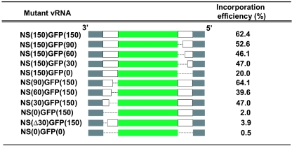
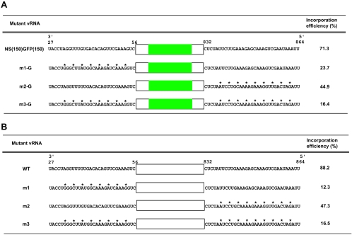
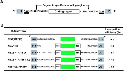
The genome of influenza A virus consists of eight single-strand negative-sense RNA segments, each comprised of a coding region and a noncoding region. The noncoding region of the NS segment is thought to provide the signal for packaging; however, we recently showed that the coding regions located at both ends of the hemagglutinin and neuraminidase segments were important for their incorporation into virions. In an effort to improve our understanding of the mechanism of influenza virus genome packaging, we sought to identify the regions of NS viral RNA (vRNA) that are required for its efficient incorporation into virions. Deletion analysis showed that the first 30 nucleotides of the 3' coding region are critical for efficient NS vRNA incorporation and that deletion of the 3' segment-specific noncoding region drastically reduces NS vRNA incorporation into virions. Furthermore, silent mutations in the first 30 nucleotides of the 3' NS coding region reduced the incorporation efficiency of the NS segment and affected virus replication. These results suggested that segment-specific noncoding regions together with adjacent coding regions (especially at the 3' end) form a structure that is required for efficient influenza A virus vRNA packaging.


Figures

FIG. 1.
FIG. 1.
Schematic representation of the system used to determine the efficiency of vRNA incorporation into virions. 293T cells were transfected with plasmids for production of influenza virus VLPs. Forty-eight hours later, aliquots of the supernatant were used to infect MDCK cells. To determine efficiency of incorporation of mutant NS vRNA segments, GFP- and NP-expressing cells were counted at 24 h postinfection.
FIG. 2.
FIG. 2.
Efficiency of incorporation of recombinant NS segments. Incorporation efficiencies of recombinant NS segments with different lengths of NS coding regions were determined as described in the legend to Fig. 1. The noncoding and coding regions of the NS vRNA are represented by grey and white bars, respectively, while the green bars represent the coding region of the GFP gene. The dotted line represents deleted sequences of the NS coding region. The results shown are representative data from three independent experiments.
FIG. 3.
FIG. 3.
Efficiencies of incorporation of NS segments with nucleotide substitutions in the NS coding regions. The nucleotide sequences at the 3′ (positions 27 to 56) or 5′ (positions 832 to 864) end of the NS coding region are shown. The silent mutations introduced are shown by asterisks. The coding regions of the NS vRNA and GFP are represented by white and green bars, respectively. The results shown are representative data for three independent experiments. (A) Efficiencies of incorporation of the NS-GFP segment with nucleotide substitutions in the NS coding regions. Incorporation efficiencies were determined as described in the legend to Fig. 1. (B) Efficiencies of incorporation of the NS segment with nucleotide substitutions in the NS coding regions. To determine the incorporation efficiency of mutant NS vRNA segments, NS2- and NP-expressing cells were counted at 24 h postinfection.
FIG. 4.
FIG. 4.
Growth properties of mutant viruses containing silent mutations in the coding regions of the NS segment. MDCK cells were infected with virus at an multiplicity of infection of 0.001. At the indicated times after infection, the virus titer in the supernatant was determined. The values are means and standard deviations from triplicate experiments. •, m1; ○, m2; ▪, m3; □, wild type.
FIG. 5.
FIG. 5.
Importance of NS segment-specific noncoding regions for incorporation of recombinant NS segments. (A) Schematic diagram of the NS vRNA. Segment-specific complementary sequences are underlined. The poly(U) sequence for the addition of the poly(A) tail to the mRNA is shown in italics. The grey boxes represent U12 and U13. (B) Efficiencies of incorporation of recombinant NS segments with deletions in the NS segment-specific noncoding regions. U12 and U13 are represented by grey boxes. The coding regions of the NS vRNA and GFP gene are represented by white and green bars, respectively. The dotted lines represent sequences deleted from the NS segment. The results shown are representative data from three independent experiments.
FIG. 6.
FIG. 6.
Comparison of test NS vRNA amounts and GFP expression levels in plasmid-transfected cells. (A) Quantitative analysis of NS(0)GFP(0), NSΔNTR, and NS(150)GFP(150) vRNAs produced from PolI plasmids in 293T cells by real-time quantitative reverse transcription-PCR. 293T cells (106 cells) were transfected with 18 plasmids. At 48 h posttransfection, total RNA was extracted from 293T cells and quantified by real-time PCR with GFP and NP sequence-specific primers and probes as described in Materials and Methods. The primers and probes for the NS-GFP segments detect NS-GFP but not the authentic NS segment. Experiments were performed in the presence (+) or absence (−) of reverse transcriptase (RT) to demonstrate that the results are not affected by the plasmids used for the experiments. NS2KO represents NS2 knockout virus, which does not produce NS2(NEP) protein and does not undergo multiple cycles of replication (27). It therefore serves as a negative control for detection of NS-GFP and a positive control for the NP segment. The results shown are representative data from three independent experiments. Error bars indicate standard deviations. (B) Comparison of GFP expression levels in PolI plasmid-transfected 293T cells by fluorescence microscopy. At 48 h after transfection, the cells were observed by fluorescence microscopy.
FIG. 7.
FIG. 7.
Sequence alignment of the NS incorporation signals among influenza A viruses. The red, orange, and purple lines represent U12, incorporation signal, and U13, respectively. The conserved sequences among all strains are shown in black. The NS sequences were obtained from the influenza A virus sequence database at Los Alamos National Laboratory. (A) Comparison of the incorporation signals among allele A NS segments. Human viruses: WSN, A/WSN/33; PR834, A/Puerto Rico/8/34; HK481, A/Hong Kong/481/97; FM47, A/Fort Monmouth/1/47; AL77, A/Alaska/6/77; SWCO77, A/swine/Colorado/1/77; BM18, A/Brevig Mission/1/18. Eurasian avian viruses: SWNE85, A/swine/Netherlands/12/85; SWCH78, A/swine/China/8/78. North American avian viruses: RTNJ85, A/ruddy turnstone/New Jersey/47/85; WHME84, A/whale/Marine/328/84; GDE471-86, A/gull/Delaware/471/86. Equine viruses: EQKY76, A/equine/Kentucky/76; EQCO76, A/equine/Cordaba/4/76. Classic swine viruses: SWIA30, A/swine/Iowa/15/30; SWIA88, A/swine/Italy/671/87. EQPR56-like equine viruses: EQPR56, A/equine/Prague/1/56; EQDET64, A/equine/Detroit/3/64. Gull viruses: GDE87, A/gull/Delaware/2838/87; GMN81, A/gull/Minnesota/1352/81. (B) Comparison of the incorporation signals among allele B NS segments. DKAL60, A/duck/Alberta/60/76; MAL827-78, A/mallard/Alberta/827/78; MAL88-76, A/mallard/Alberta/88/76; PAL121-79, A/pintail/Alberta/121/79; PAL358-79, A/pintail/Alberta/358/79. The sequence data for BM18, MAL827-78, MAL88-76, PAL121-79, and PAL358-79 are not complete in the influenza virus database.

References

    1. Baez, M., J. J. Zazra, R. M. Elliott, J. F. Young, and P. Palese. 1981. Nucleotide sequence of the influenza A/duck/Alberta/60/76 virus NS RNA: conservation of the NS1/NS2 overlapping gene structure in a divergent influenza virus RNA segment. Virology 113:397-402. - PubMed
    1. Bancroft, C. T., and T. G. Parslow. 2002. Evidence for segment-nonspecific packaging of the influenza A virus genome. J. Virol. 76:7133-7139. - PMC - PubMed
    1. Chen, W., P. A. Calvo, D. Malide, J. Gibbs, U. Schubert, I. Bacik, S. Basta, R. O'Neill, J. Schickli, P. Palese, P. Henklein, J. R. Bennink, and J. W. Yewdell. 2001. A novel influenza A virus mitochondrial protein that induces cell death. Nat. Med. 7:1306-1312. - PubMed
    1. Elton, D., M. Simpson-Holley, K. Archer, L. Medcalf, R. Hallam, J. McCauley, and P. Digard. 2001. Interaction of the influenza virus nucleoprotein with the cellular CRM1-mediated nuclear export pathway. J. Virol. 75:408-419. - PMC - PubMed
    1. Enami, M., R. Fukuda, and A. Ishihama. 1985. Transcription and replication of eight RNA segments of influenza virus. Virology 142:68-77. - PubMed

Publication types

MeSH terms

LinkOut - more resources