

doi: 10.1128/JVI.79.6.3841-3845.2005.
Deletion of the second immunoglobulin-like domain of nectin-1 alters its intracellular processing and localization and ability to mediate entry of herpes simplex virus
Affiliations
- PMID: 15731277
- PMCID: PMC1075719
- DOI: 10.1128/JVI.79.6.3841-3845.2005
Item in Clipboard
Deletion of the second immunoglobulin-like domain of nectin-1 alters its intracellular processing and localization and ability to mediate entry of herpes simplex virus
J Virol.
2005 Mar.
Abstract
Nectin-1 is an immunoglobulin (Ig)-like entry receptor for herpes simplex virus (HSV). Like other nectins, nectin-1 forms dimers and mediates cell adhesion through interactions with other nectins. We constructed a second-domain deletion mutant of nectin-1 (nectin-1-Delta2) to examine the role of the second Ig-like domain in HSV entry. Nectin-1-Delta2 exhibited a severely reduced ability to mediate HSV entry and accumulated in the endoplasmic reticulum but retained the ability to interact with its HSV ligand, gD. The failure of nectin-1-Delta2 to mediate HSV entry probably resulted from its failure to be transported to a membrane targeted by HSV for viral entry.
Figures
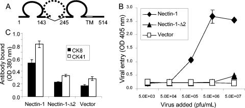
Structure, HSV entry activity, and cell surface expression of nectin-1-Δ2. (A) Schematic representation of the nectin-1-Δ2 mutant, with amino acid numbering (based on GenBank reference sequence NP_002846) shown below. (B) Nectin-1-Δ2 does not mediate entry of HSV-1. CHO-K1 cells transfected with receptor or vector control were grown to confluency in 96-well tissue culture plates and exposed to increasing doses of reporter virus HSV-1(KOS)tk12 that expresses β-galactosidase upon viral entry (23). After 6 h of incubation at 37°C, cells were permeabilized and ONPG substrate was added. Viral entry was quantified by measuring optical densities at 405 nm. Error bars represent 1 standard deviation of triplicate determinations within the same experiment, and the results shown here are representative of three independent experiments. (C) Cell surface expression of nectin-1 and nectin-1-Δ2. CHO-K1 cells were transfected with plasmids expressing nectin-1, nectin-1-Δ2, or vector control, replated in 96-well plates, and then incubated with anti-nectin-1 monoclonal antibodies CK8 or CK41. Following washes to remove unbound antibodies, the cells were fixed and incubated with secondary antibodies and the horseradish peroxidase detection system described elsewhere (6). Results of the cell-based enzyme-linked immunosorbent assays are expressed as optical density (OD) at 380 nm and are means of triplicate measurements, with error bars indicating 1 standard deviation. The results shown are representative of three independent experiments.
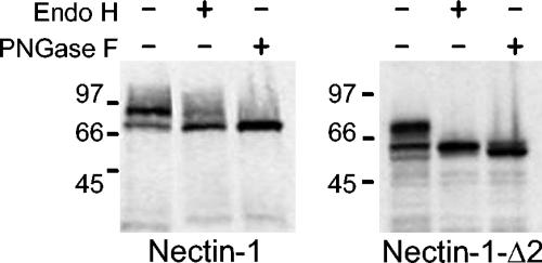
Western blot analysis of nectin-1 and nectin-1-Δ2 after endoglycosidase treatment. CHO-K1 cells were transfected with tagged versions of nectin-1 and nectin-1-Δ2. The cells were lysed 48 h after transfection, cell lysates were treated with Endo H or PNGase F and run on a denaturing SDS-PAGE gel, and the expressed proteins were visualized by Western blotting using antibodies specific for the tags.
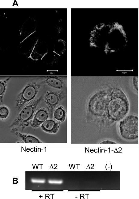
Cellular localization and expression of nectin-1 and nectin-1-Δ2. (A) CHO-K1 cells were transfected with plasmids expressing nectin-1 or nectin-1-Δ2 and stained with anti-nectin-1 antibody (top panels) or visualized by phase-contrast microscopy (bottom panels). Nectin-1 localized to cell-cell junctions, whereas nectin-1-Δ2 expression was mainly confined to the cytoplasm. Bars, 10 μm. (B) Similar amounts of nectin-1 (WT) and nectin-1-Δ2 (Δ2) mRNA were detected by reverse transcriptase PCR using primers that amplify a 247-bp region upstream of the deletion in nectin-1-Δ2. Total RNA extracted from transfected CHO cells using the RNeasy kit (QIAGEN) was subjected to DNase I treatment (Invitrogen). RNA samples before (-RT) or after (+RT) reverse transcription (Promega) using an oligo(dT) primer were used as templates in the PCR. (-), negative control (water).
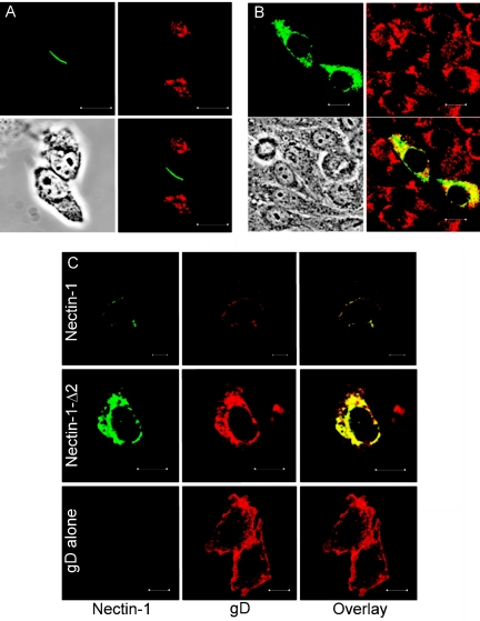
Costaining of wild-type and mutant nectin-1 with calreticulin or viral gD. (A) Nectin-1 does not colocalize with calreticulin in CHO cells transfected with nectin-1. (B) Nectin-1-Δ2 colocalizes with calreticulin in CHO cells transfected with nectin-1-Δ2. Primary antibodies used in panels A and B were chicken anti-nectin-1 (green) and rabbit anti-calreticulin (red; Abcam). Bottom right of each panel shows the overlay, and the bottom left shows a phase-contrast image. (C) Colocalization of HSV gD and wild-type or mutant nectin-1. CHO-K1 cells were transfected with plasmid pCJ3, expressing HSV-1 gD (6), or cotransfected with plasmids expressing HSV-1 gD and either nectin-1 or nectin-1-Δ2, stained with chicken anti-nectin-1 antibody (green) and rabbit anti-gD antibody R7 (red), and visualized by confocal microscopy. As in cells transfected with receptor alone, nectin-1 localized to cell-cell junctions, whereas nectin-1-Δ2 localized to the cytoplasm. HSV-1 gD was mostly colocalized with nectin-1 or nectin-1-Δ2 but, when expressed alone, had a different intracellular localization than when expressed with either form of nectin-1. Bars, 10 μm.
Similar articles
-
Use of chimeric nectin-1(HveC)-related receptors to demonstrate that ability to bind alphaherpesvirus gD is not necessarily sufficient for viral entry.Virology. 2001 Jul 5;285(2):366-75. doi: 10.1006/viro.2001.0989. Virology. 2001. PMID: 11437670
-
Contributions of gD receptors and glycosaminoglycan sulfation to cell fusion mediated by herpes simplex virus 1.Virus Res. 2001 Apr;74(1-2):39-45. doi: 10.1016/s0168-1702(00)00244-6. Virus Res. 2001. PMID: 11226572
-
Structural features of nectin-2 (HveB) required for herpes simplex virus entry.J Virol. 2001 Nov;75(22):11185-95. doi: 10.1128/JVI.75.22.11185-11195.2001. J Virol. 2001. PMID: 11602758 Free PMC article.
-
Different receptors binding to distinct interfaces on herpes simplex virus gD can trigger events leading to cell fusion and viral entry.Virology. 2006 Jan 5;344(1):17-24. doi: 10.1016/j.virol.2005.09.016. Virology. 2006. PMID: 16364731 Review.
-
The novel receptors that mediate the entry of herpes simplex viruses and animal alphaherpesviruses into cells.Rev Med Virol. 2000 Sep-Oct;10(5):305-19. doi: 10.1002/1099-1654(200009/10)10:5<305::aid-rmv286>3.0.co;2-t. Rev Med Virol. 2000. PMID: 11015742 Review.
Cited by
-
Novel mutations in gB and gH circumvent the requirement for known gD Receptors in herpes simplex virus 1 entry and cell-to-cell spread.J Virol. 2013 Feb;87(3):1430-42. doi: 10.1128/JVI.02804-12. Epub 2012 Nov 14. J Virol. 2013. PMID: 23152509 Free PMC article.
-
A soluble form of human nectin-2 impairs exocrine secretion of pancreas and formation of zymogen granules in transgenic mice.Biochem Biophys Rep. 2015 Dec 10;5:196-202. doi: 10.1016/j.bbrep.2015.12.006. eCollection 2016 Mar. Biochem Biophys Rep. 2015. PMID: 28955824 Free PMC article.
-
Herpes virus fusion and entry: a story with many characters.Viruses. 2012 May;4(5):800-32. doi: 10.3390/v4050800. Epub 2012 May 10. Viruses. 2012. PMID: 22754650 Free PMC article. Review.
-
Herpes B virus utilizes human nectin-1 but not HVEM or PILRα for cell-cell fusion and virus entry.J Virol. 2012 Apr;86(8):4468-76. doi: 10.1128/JVI.00041-12. Epub 2012 Feb 15. J Virol. 2012. PMID: 22345445 Free PMC article.
-
The Ig-like v-type domain of paired Ig-like type 2 receptor alpha is critical for herpes simplex virus type 1-mediated membrane fusion.J Virol. 2010 Sep;84(17):8664-72. doi: 10.1128/JVI.01039-10. Epub 2010 Jun 23. J Virol. 2010. PMID: 20573830 Free PMC article.
References
-
- Cocchi, F., M. Lopez, L. Menotti, M. Aoubala, P. Dubreuil, and G. Campadelli-Fiume. 1998. The V domain of herpesvirus Ig-like receptor (HIgR) contains a major functional region in herpes simplex virus-1 entry into cells and interacts physically with the viral glycoprotein D. Proc. Natl. Acad. Sci. USA 95:15700-15705. - PMC - PubMed
-
- Cocchi, F., L. Menotti, P. Mirandola, M. Lopez, and G. Campadelli-Fiume. 1998. The ectodomain of a novel member of the immunoglobulin subfamily related to the poliovirus receptor has the attributes of a bona fide receptor for herpes simplex virus types 1 and 2 in human cells. J. Virol. 72:9992-10002. - PMC - PubMed
-
- Fabre, S., N. Reymond, F. Cocchi, L. Menotti, P. Dubreuil, G. Campadelli-Fiume, and M. Lopez. 2002. Prominent role of the Ig-like V domain in trans-interactions of nectins. Nectin3 and nectin 4 bind to the predicted C-C′-C"-D beta-strands of the nectin1 V domain. J. Biol. Chem. 277:27006-27013. - PubMed
-
- Geraghty, R. J., A. Fridberg, C. Krummenacher, G. H. Cohen, R. J. Eisenberg, and P. G. Spear. 2001. Use of chimeric nectin-1(HveC)-related receptors to demonstrate that ability to bind alphaherpesvirus gD is not necessarily sufficient for viral entry. Virology 285:366-375. - PubMed
Publication types
MeSH terms
Substances
Grants and funding
LinkOut - more resources
Full Text Sources
Molecular Biology Databases
Miscellaneous
