

Exogenous ACE2 expression allows refractory cell lines to support severe acute respiratory syndrome coronavirus replication
- PMID: 15731278
- PMCID: PMC1075706
- DOI: 10.1128/JVI.79.6.3846-3850.2005
Exogenous ACE2 expression allows refractory cell lines to support severe acute respiratory syndrome coronavirus replication
Abstract
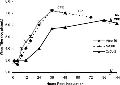
Of 30 cell lines and primary cells examined, productive severe acute respiratory syndrome coronavirus (Urbani strain) (SARS-CoV) infection after low-multiplicity inoculation was detected in only six: three African green monkey kidney epithelial cell lines (Vero, Vero E6, and MA104), a human colon epithelial line (CaCo-2), a porcine kidney epithelial line [PK(15)], and mink lung epithelial cells (Mv 1 Lu). SARS-CoV produced a lytic infection in Vero, Vero E6, and MA104 cells, but there was no visible cytopathic effect in Caco-2, Mv 1 Lu, or PK(15) cells. Multistep growth kinetics were identical in Vero E6 and MA104 cells, with maximum titer reached 24 h postinoculation (hpi). Virus titer was maximal 96 hpi in CaCo-2 cells, and virus was continually produced from infected CaCo-2 cells for at least 6 weeks after infection. CaCo-2 was the only human cell type of 13 tested that supported efficient SARS-CoV replication. Expression of the SARS-CoV receptor, angiotensin-converting enzyme 2 (ACE2), resulted in SARS-CoV replication in all refractory cell lines examined. Titers achieved were variable and dependent upon the method of ACE2 expression.
Figures
References
Publication types
MeSH terms
Substances
Grants and funding
LinkOut - more resources
Full Text Sources
Other Literature Sources
Research Materials
Miscellaneous
