. 2005 Mar;79(6):3865-72.
doi: 10.1128/JVI.79.6.3865-3872.2005.

Endocytosis of the Nipah virus glycoproteins

Affiliations

Endocytosis of the Nipah virus glycoproteins

Carola Vogt et al. J Virol. 2005 Mar.

Abstract

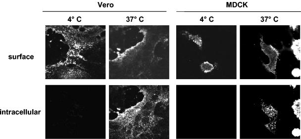
Nipah virus (NiV), a highly pathogenic member of the family Paramyxoviridae, encodes the surface glycoproteins F and G. Since internalization of the NiV envelope proteins from the cell surface might be of functional importance for viral pathogenesis either by regulating cytopathogenicity or by modulating recognition of infected cells by the immune system, we analyzed the endocytosis of the NiV F and G proteins. Interestingly, we found both glycoproteins to be internalized in infected and transfected cells. As endocytosis is normally mediated by tyrosine- or dileucine-dependent signals in the cytoplasmic tails of transmembrane proteins, all potential internalization signals in the NiV glycoproteins were mutated. Whereas the G protein appeared to be constitutively internalized with the bulk flow during membrane turnover, uptake of the F protein was found to be signal mediated. F endocytosis clearly depended on a membrane-proximal YXXPhi motif and was found to be of functional importance for the biological activity of the protein.


Figures

FIG. 1.
FIG. 1.
Confocal microscopy analysis of NiV glycoprotein endocytosis in infected cells. Vero and MDCK cells were infected with NiV at a multiplicity of infection of 0.1. At 24 h p.i., cells were incubated with an NiV-specific guinea pig antiserum at 4°C for 30 min without prior fixation. Then, cells were either kept on ice (4°C) or warmed to 37°C to allow endocytosis to occur (37°C). After 30 min, cells were fixed with 4% paraformaldehyde for 16 h, and surface-bound antibodies were detected by incubation with an FITC-conjugated secondary antibody (surface). After permeabilization of the cells with methanol-acetone, internalized antibodies were stained with a rhodamine-conjugated secondary antibody (intracellular). The images shown represent the central sections from the cells analyzed by laser scanning confocal microscopy.
FIG. 2.
FIG. 2.
Endocytosis of singly expressed or coexpressed NiV F and G proteins. (A) MDCK cells were transfected with pczCFG5 plasmids encoding either the NiV F or G proteins or were cotransfected with both plasmids (F + G). At 24 h posttransfection, the cells were incubated with the NiV-specific antiserum at 4°C for 30 min and then warmed to 37°C to allow endocytosis to occur. Surface-bound antibodies were visualized by FITC-conjugated secondary antibodies (surface), and internalized primary antibodies were stained after permeabilization with methanol-acetone by rhodamine-conjugated secondary antibodies (intracellular). (B) MDCK cells expressing F, G, or F and G (F+G) proteins were surface labeled with cleavable NHS-SS-biotin at 4°C. Then, the cells were warmed to 37°C for the times indicated to allow endocytosis of the biotinylated proteins to occur. Subsequently, cell surface proteins were reduced with MESNA. To determine the total amount of surface-biotinylated proteins, the cells were compared to cells that were neither warmed to 37°C nor treated with MESNA (control). After cell lysis, F and G proteins were immunoprecipitated with the NiV-specific antiserum. The samples were separated on a sodium dodecyl sulfate-10% polyacrylamide gel under nonreducing conditions and transferred to nitrocellulose. Biotinylated proteins were then detected with peroxidase-conjugated streptavidin. The control lane represents 50% of the total amount of biotinylated F or G proteins. (C) To determine the rate of internalization, the percentages of internalized F (♦) and G (•) proteins measured in the experiment shown in Fig. 2B (panels F and G) were plotted as a function of the time that cells were incubated at 37°C.
FIG. 3.
FIG. 3.
Internalization of F and G proteins with mutations in the potential endocytosis signals. (A) Sequences of the cytoplasmic domains of wild-type and mutant G and F proteins. The numbers above the sequences indicate the amino acid positions. Amino acid sequences are shown in a single-letter code; boldface letters indicate exchanged amino acid residues. The vertical lines separate the transmembrane domains from those predicted to be in the cytoplasm. (B and C) Antibody uptake analysis of mutant G (GY28/29A and GL41/42A) (B) and mutant F (FY542/543A, FY525A, and FYA) (C) proteins was performed as described in the legend to Fig. 2A. (D) Endocytosis of mutant F proteins (FY542/543A, FY525A, and FYA) was determined after surface biotinylation and MESNA reduction as described in the legend to Fig. 2B. (E) The rates of internalization were determined by plotting the percentages of endocytosed FY542/543A (⧫), FY525A (•), and FYA (▴) proteins as functions of the times that the cells were incubated at 37°C. (F) The total amount of surface-expressed wild-type (F) and mutant F (FY542/543A, FY525A, and FYA) proteins was determined by the surface biotinylation of 2 × 10⁶ cells at 30 h posttransfection. After cell lysis, the F proteins were immunoprecipitated and detected as described in the legend to Fig. 2A.
FIG. 3.
FIG. 3.
Internalization of F and G proteins with mutations in the potential endocytosis signals. (A) Sequences of the cytoplasmic domains of wild-type and mutant G and F proteins. The numbers above the sequences indicate the amino acid positions. Amino acid sequences are shown in a single-letter code; boldface letters indicate exchanged amino acid residues. The vertical lines separate the transmembrane domains from those predicted to be in the cytoplasm. (B and C) Antibody uptake analysis of mutant G (GY28/29A and GL41/42A) (B) and mutant F (FY542/543A, FY525A, and FYA) (C) proteins was performed as described in the legend to Fig. 2A. (D) Endocytosis of mutant F proteins (FY542/543A, FY525A, and FYA) was determined after surface biotinylation and MESNA reduction as described in the legend to Fig. 2B. (E) The rates of internalization were determined by plotting the percentages of endocytosed FY542/543A (⧫), FY525A (•), and FYA (▴) proteins as functions of the times that the cells were incubated at 37°C. (F) The total amount of surface-expressed wild-type (F) and mutant F (FY542/543A, FY525A, and FYA) proteins was determined by the surface biotinylation of 2 × 10⁶ cells at 30 h posttransfection. After cell lysis, the F proteins were immunoprecipitated and detected as described in the legend to Fig. 2A.
FIG. 4.
FIG. 4.
Biological activity of mutant G and F proteins. MDCK cells were cotransfected with plasmids encoding either wild-type or mutant G or F proteins as indicated. To visualize syncytium formation, the cells were fixed at 24 h posttransfection with ethanol and stained with Giemsa staining solution. Representative microscopic fields were photographed.

Similar articles

Cited by

References

    1. Berlioz-Torrent, C., B. L. Shacklett, L. Erdtmann, L. Delamarre, I. Bouchaert, P. Sonigo, M. C. Dokhelar, and R. Benarous. 1999. Interactions of the cytoplasmic domains of human and simian retroviral transmembrane proteins with components of the clathrin adaptor complexes modulate intracellular and cell surface expression of envelope glycoproteins. J. Virol. 73:1350-1361. - PMC - PubMed
    1. Bonifacino, J. S., and E. C. Dell'Angelica. 1999. Molecular bases for the recognition of tyrosine-based sorting signals. J. Cell Biol. 145:923-926. - PMC - PubMed
    1. Bonifacino, J. S., and L. M. Traub. 2003. Signals for sorting of transmembrane proteins to endosomes and lysosomes. Annu. Rev. Biochem. 72:395-447. - PubMed
    1. Bossart, K. N., L.-F. Wang, M. N. Flora, K. B. Chua, S. K. Lam, B. T. Eaton, and C. C. Broder. 2002. Membrane fusion tropism and heterotypic functional activities of the Nipah virus and Hendra virus envelope glycoproteins. J. Virol. 76:11186-11198. - PMC - PubMed
    1. Chua, K. B., K. J. Goh, K. T. Wong, A. Kamarulzaman, P. S. Tan, T. G. Ksiazek, S. R. Zaki, G. Paul, S. K. Lam, and C. T. Tan. 1999. Fatal encephalitis due to Nipah virus among pig-farmers in Malaysia. Lancet 354:1257-1259. - PubMed

Publication types

MeSH terms