. 2005 Feb 24;33(4):1182-92.
doi: 10.1093/nar/gki257. Print 2005.

The effect of chemical modifications on the thermal stability of different G-quadruplex-forming oligonucleotides

Affiliations

The effect of chemical modifications on the thermal stability of different G-quadruplex-forming oligonucleotides

Barbara Saccà et al. Nucleic Acids Res. .

Abstract

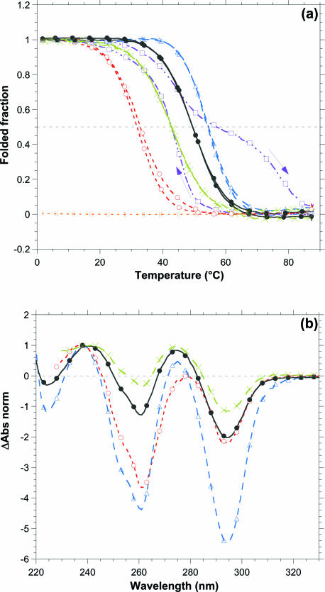
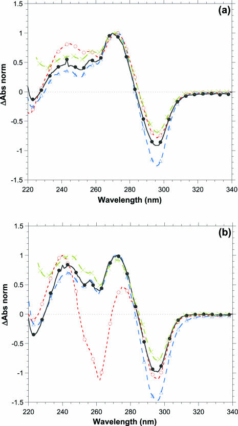
A systematic study of the thermal and conformational properties of chemically modified G-quadruplexes of different molecularities is reported. The effect of backbone charge and atom size, thymine/uracyl substitution as well as the effect of modification at the ribose 2'-position was analyzed by UV spectroscopy. Additional calorimetric studies were performed on different modified forms of the human telomeric sequence. Determination of the differential spectra allowed more insights into the conformational properties of the oligonucleotides. Lack of negative charge at the phosphate backbone yielded to a general destabilization of the G-quadruplex structure. On the other hand, substitution of thymine with uracyl resulted in a moderate or strong stabilization of the structure. Additional modification at the sugar 2'-position gave rise to different effects depending on the molecularity of the quadruplex. In particular, loss of hydrogen bond capacity at the 2'-position strongly affected the conformation of the G-quadruplex. Altogether, these results demonstrate that the effect of some modifications depends on the sequence context, thus providing helpful information for the use of chemically modified quadruplexes as therapeutic agents or as structural elements of supramolecular complexes.


Figures

Figure 1
Figure 1
(a) Structure of a G-quartet: cyclic array of four guanines, linked by Hoogsten hydrogen bonds and stabilized by an internal positive ion. (b) Schematic representation of different G-quadruplex structures, with variable number and relative orientation of the self-associated strands and different orientation of the loops.
Figure 2
Figure 2
Schematic representation of the chemical modifications examined in this work and nomenclature used to designate the different modified oligonucleotides. The backbone modified oligos are indicated as oligo-S for the phosphorothioate analogs and oligo-M for the methylphosphonate analogs. The deoxyribonucleotide derivatives (T→U substitution only) are indicated as oligo-U. Additional modification at the sugar level is indicated as oligo-R for the ribonucleotide analogs or oligo-O for the 2′-O-methyl-ribonucleotide analogs.
Figure 3
Figure 3
(a) Normalized thermal transition profiles measured at 295 nm and (b) normalized differential spectra in the 220–340 nm region for the 15TBA oligo and its analogs at 5 μM (or 3 μM for 15TBA-S) strand concentration in 10 mM sodium cacodylate buffer pH 7.0 containing 100 mM KCl. 15TBA (black full circles), 15TBA-S (green crosses), 15TBA-M (yellow vertical bars), 15TBA-R (violet open squares), 15TBA-O (red open circles) and 15TBA-U (blue open triangles). An hysteresis phenomenon occurs for the 15TBA-R and the direction of the temperature gradient is indicated by the arrows.
Figure 4
Figure 4
Normalized differential spectra in the 220–340 nm region for the 22AG oligo and its S-, O- and U-analogs at 5 μM strand concentration in 10 mM sodium cacodylate buffer pH 7.0 containing (a) 100 mM NaCl or (b) 100 mM KCl. 22AG (black full circles), 22AG-S (green crosses), 22AG-O (red open circles) and 22AG-U (blue open triangles).
Figure 5
Figure 5
Normalized thermal denaturation profiles measured at 295 nm and normalized differential spectra in the 220–340 nm region for the 12G4 series [(a) and (b), respectively] and TG4 series [(c) and (d), respectively] at 10 μM strand concentration in 10 mM sodium cacodylate buffer, pH 7.0, containing 100 mM NaCl. 12G4 and TG4, black full circles; 12G4-S and TG4-S, green crosses; 12G4-M and TG4-M, yellow vertical bars; 12G4-R and TG4-R, violet open squares; 12G4-O and TG4-O, red open circles; and 12G4-U and TG4-U, blue open triangles. Upon cooling, no renaturation of the TG4 quadruplexes is obtained, and further heating/cooling cycles led to a similar monotonous variation of absorbance with no evidence for quadruplex reformation or denaturation.

References

    1. Keniry M. Quadruplex structures in nucleic acids. Biopolymers. 2001;56:123–146. - PubMed
    1. Neidle S., Parkinson G.N. The structure of telomeric DNA. Curr. Opin. Struct. Biol. 2003;13:275–283. - PubMed
    1. Gellert M., Lipsett M.N., Davies D.R. Helix formation by guanylic acid. Proc. Natl Acad. Sci. USA. 1962;48:2013–2018. - PMC - PubMed
    1. Blackburn E.H. Telomeres: no end in sight. Cell. 1994;77:621–623. - PubMed
    1. Sen D., Gilbert W. Formation of parallel four-stranded complexes by guanine-rich motifs in DNA and its implications for meiosis. Nature. 1988;334:364–366. - PubMed

Publication types