. 2005 Feb 24;33(4):1193-200.
doi: 10.1093/nar/gki263. Print 2005.

Separation of mutation avoidance and antirecombination functions in an Escherichia coli mutS mutant

Affiliations

Separation of mutation avoidance and antirecombination functions in an Escherichia coli mutS mutant

Melissa A Calmann et al. Nucleic Acids Res. .

Abstract

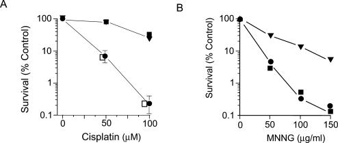
DNA mismatch repair in Escherichia coli has been shown to be involved in two distinct processes: mutation avoidance, which removes potential mutations arising as replication errors, and antirecombination which prevents recombination between related, but not identical (homeologous), DNA sequences. We show that cells with the mutSDelta800 mutation (which removes the C-terminal 53 amino acids of MutS) on a multicopy plasmid are proficient for mutation avoidance. In interspecies genetic crosses, however, recipients with the mutSDelta800 mutation show increased recombination by up to 280-fold relative to mutS+. The MutSDelta800 protein binds to O6-methylguanine mismatches but not to intrastrand platinated GG cross-links, explaining why dam bacteria with the mutSDelta800 mutation are resistant to cisplatin, but not MNNG, toxicity. The results indicate that the C-terminal end of MutS is necessary for antirecombination and cisplatin sensitization, but less significant for mutation avoidance. The inability of MutSDelta800 to form tetramers may indicate that these are the active form of MutS.


Figures

Figure 1
Figure 1
Gal reversion assay. Dark Gal+ revertants are shown on a background of white Gal colonies. From left to right: the GM4799 strain (mutS null mutation) with plasmids bearing mutS+, mutSΔ680 and mutSΔ800, respectively.
Figure 2
Figure 2
Survival of cells exposed to cisplatin and MNNG. Cell survival of strain GM5556 (Δdam-16::Kan mutS::Tn10) containing plasmids with mutS+ (closed circles), mutSΔ680 (inverted triangles), and mutSΔ800 (closed squares) after exposure to cisplatin (A) and MNNG (B). The open squares in (A) represent the survival of GM3819 (Δdam-16::Kan mutS+) with the mutSΔ800 plasmid.
Figure 3
Figure 3
Binding isotherm of MutS and MutSΔ800 to heteroduplex DNA. The upper panel shows the binding of MutS and MutSΔ800 to a P32-labeled heteroduplex DNA with a single base IDL. The heteroduplex concentration was 0.6 pmol. The data are shown in the lower panel as a binding isotherm.
Figure 4
Figure 4
Binding of MutS and MutSΔ800 to modified DNA. (A) The binding of MutS and MutSΔ800 to unplatinated (lanes 1–5) and platinated homoduplex (lanes 6–10) DNA. No enzyme (lanes 1 and 6); MutS, 8 μM (lanes 2 and 7) and 4 μM (lanes 3 and 8); and MutSΔ800, 8 μM (lanes 4 and 9) and 4 μM (lanes 5 and 10). The DNA was visualized by staining the gel with Vistra Green. (B) The binding of MutS and MutSΔ800 to unplatinated (lanes 1–5) and platinated heteroduplex (lanes 6–10). Protein additions are the same as in (A). (C) The binding of MutS and MutSΔ800 to O6-meG–C (lanes 1–4) and O6-meG–T (lanes 5–8) DNA. MutS, 8 μM (lanes 1 and 5) and 16 μM (lanes 2 and 6); and MutSΔ800, 8 μM (lanes 3 and 7) and 16 μM (lanes 4 and 8).
Figure 5
Figure 5
RecA-catalyzed strand transfer. The upper panel shows a schematic diagram of the reaction. Single-strand (SS) circular DNA reacts with linear (L) duplex to form intermediate (I) structures which are converted into nicked-circle (NC) products. The fluorograph shows the results from the homologous M13–M13 (lanes a–e) and the homeologous M13-fd (lanes f–j) reactions. Samples were removed at time 0 (lanes a and f), 5 min (lanes b and g), 15 min (lanes c and h), 45 min (lanes d and i) and 90 min (lanes e and j).
Figure 6
Figure 6
Kinetics of MutS inhibition of RecA-mediated strand exchange. The graphs show the effect of varying concentrations of MutS (A) and MutSΔ800 (B) on RecA strand exchange using homologous (M13–M13) and homeologous (M13-fd). Closed circles, M13–M13; crosses, M13–M13 plus 100 nM protein; closed squares, M13-fd; open squares, M13-fd plus 25 nM protein; open circles, M13-fd plus 100 nM protein; and inverted triangles, M13-fd plus 25 nM protein plus 40 nM MutL.

References

    1. Modrich P., Lahue R. Mismatch repair in replication fidelity, genetic recombination, and cancer biology. Annu. Rev. Biochem. 1996;65:101–133. - PubMed
    1. Schofield M.J., Hsieh P. DNA mismatch repair: molecular mechanisms and biological function. Annu. Rev. Microbiol. 2003;57:579–608. - PubMed
    1. Marti T.M., Kunz C., Fleck O. DNA mismatch repair and mutation avoidance pathways. J. Cell Physiol. 2002;191:28–41. - PubMed
    1. Rayssiguier C., Thaler D.S., Radman M. The barrier to recombination between Escherichia coli and Salmonella typhimurium is disrupted in mismatch-repair mutants. Nature. 1989;342:396–401. - PubMed
    1. Westmoreland J., Porter G., Radman M., Resnick M.A. Highly mismatched molecules resembling recombination intermediates efficiently transform mismatch repair proficient Escherichia coli. Genetics. 1997;145:29–38. - PMC - PubMed

Publication types

MeSH terms