. 2005 Feb 24;33(4):1240-8.
doi: 10.1093/nar/gki278. Print 2005.

Target-dependent on/off switch increases ribozyme fidelity

Affiliations

Target-dependent on/off switch increases ribozyme fidelity

Lucien Junior Bergeron et al. Nucleic Acids Res. .

Abstract

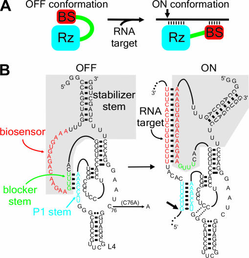
Ribozymes, RNA molecules that catalyze the cleavage of RNA substrates, provide an interesting alternative to the RNA interference (RNAi) approach to gene inactivation, especially given the fact that RNAi seems to trigger an immunological response. Unfortunately, the limited substrate specificity of ribozymes is considered to be a significant hurdle in their development as molecular tools. Here, we report the molecular engineering of a ribozyme possessing a new biosensor module that switches the cleavage activity from 'off' (a 'safety lock') to 'on' solely in the presence of the appropriate RNA target substrate. Both proof-of-concept and the mechanism of action of this man-made riboswitch are demonstrated using hepatitis delta virus ribozymes that cleave RNA transcripts derived from the hepatitis B and C viruses. To our knowledge, this is the first report of a ribozyme bearing a target-dependent module that is activated by its RNA substrate, an arrangement which greatly diminishes non-specific effects. This new approach provides a highly specific and improved tool with significant potential for application in the fields of both functional genomics and gene therapy.


Figures

Figure 1
Figure 1
The concept of the SOFA-ribozyme. (A) Schematic representation of both the ‘off’ and ‘on’ conformations of the SOFA-ribozyme. The ribozyme (Rz) and the biosensor (BS) are in blue and red, respectively. The small arrow indicates the cleavage site. (B) Secondary structure and nucleotide sequence of SOFA+-δRz-303 in both the ‘off’ and the ‘on’ conformations. The gray section indicates the SOFA module. The P1 stem of the ribozyme and the biosensor are in blue and red, respectively. The blocker sequence is represented in green. The C76A mutation that produces an inactive ribozyme version is indicated. The numbering system is according to (15).
Figure 2
Figure 2
Rational design and proof-of-concept of the SOFA-ribozyme. (A) Schematic representation and typical autoradiogram of a denaturing 6% PAGE gel used to analyze the cleavage reactions of various versions of ribozyme. The P1 stem of the ribozyme (Rz), the blocker and the biosensor (BS) are in blue, green and red, respectively. The length of the bands, in nucleotides, is shown adjacent to the gel. The control (−) was performed in the absence of ribozyme (lane1), while lane 2 is in the presence of the original δRz. Lanes 3–5 are ribozymes progressively extended by the addition of the blocker (lane 3), the biosensor (lane 4) and the stabilizer (lane 5). Lane 6 is a SOFA-ribozyme that consists of a ribozyme harboring an inappropriate biosensor sequence. (B) Typical autoradiogram of a denaturing 6% PAGE gel used to analyzing the cleavage reaction of the HBV-derived target by the original- (WT), SOFA-δRz-303 and -513 ribozymes. The incubation time was 3 h at 37°C. The length of the bands in nucleotides is shown adjacent to the gel. The control (−) was performed in the absence of ribozyme, while SOFA+ and SOFA indicates SOFA harboring either the appropriate or inappropriate biosensor sequences, respectively. (C) Graphical representations of time courses for the cleavage reactions of SOFA+ (squares), SOFA (circles) and the original (inversed triangles) ribozyme versions of δRz-303 (filled symbols) and δRz-513 (open symbols). Incubations were performed at 37°C and aliquots were recovered at 0, 2, 5, 10, 15, 20, 40, 60, 120 and 180 min.
Figure 3
Figure 3
Characterization of SOFA+-δRz-303. (A) Autoradiogram of a denaturing 6% PAGE gel showing the cleavage assays of the HBV-derived substrate by SOFA+-δRz-303 bearing biosensor stems of various lengths (BS-X, where X indicates the length of the stem). The reactions were performed under single turnover conditions at 37°C for 3 h. (B) Autoradiogram of a 20% PAGE gel showing the cleavage assays of the 44 nt HBV-derived substrate by a SOFA+-δRz-303 bearing a biosensor stem of various lengths. The reactions were performed at 37°C for 2 h under multiple turnover conditions. XC indicates the position of the xylene cyanol dye. For (A) and (B), the length of the bands in nucleotides is shown adjacent to the gel. The control (−) was performed in the absence of ribozyme.
Figure 4
Figure 4
Analysis of the mechanism of action of the SOFA-δ ribozyme. The upper panel shows the proposed sequential interactions between the δRz and the substrate. The roman numerals identify the steps of the mechanism. Dashed lines indicate oligonucleotide binding to either the substrate (i.e. FCO acting as facilitator) or the biosensor (BSO). The lower panel is a histogram showing the relative cleavage efficiencies as calculated from two independent sets of experiments using the original- (WT), SOFA+- and SOFA-δRz-303 ribozymes incubated either with or without the FCO, BSO and unrelated (UNO) oligonucleotides.
Figure 5
Figure 5
Trans-cleavage analysis of various SOFA-δ ribozymes. (A) Schematic representation of the HBV target substrates. All the ribozymes and substrates have a similar P1 stem sequences (in blue), but differ in their biosensor sequences (in various colors). The small arrow indicates the cleavage site (for the sequences, see Supplementary Table 1). (B) Cleavage assays of a pool of ten 5′ end-labeled substrates (a–j) by either a specific SOFA-δ ribozyme (named A–J), or the original ribozyme (WT). The reactions were performed at 37°C for 3 h. BPB and XC indicate the positions of the bromophenol blue and xylene cyanol dyes, respectively. The control (−) was performed in the absence of ribozyme.
Figure 6
Figure 6
Cis-cleavage analysis of various SOFA-δ ribozymes. (A and C) Schematic representation of the HBV and HCV target substrates, respectively. For these experiments, the sequence of the P1 stem is identical at each site, but the biosensor sequences are different (see the legend on the left of the Figure). (B) Autoradiogram of the cleavage assays of the 1190 nt HBV-derived target by SOFA-ribozymes cleaving at either position 398 or 993. Lane 1 is the control performed in the absence of ribozyme. Lane 2 is the reaction performed in the presence of the original δRz that cleaves at both sites (positions 398 and 993). Lanes 3 and 4 are the reactions performed with the SOFA+-δRz-398 and −993 ribozymes, respectively. Finally, lane 5 is the reaction performed in the presence of both the SOFA+-δRz-398 and -993. The reactions were performed at 37°C for 3 h. (D) Autoradiogram of the cleavage assays of a 1422 nt HCV-derived target by SOFA-δ ribozymes cleaving at either position 224 or 302 of the IRES. Lane 1 is the control performed in the absence of ribozyme. Lane 2 is the reaction performed in the presence of the original δRz that cleaves at both sites (positions 224 and 302). Lanes 3 and 4 are the reactions performed with the SOFA+-δRz-224 and -302 ribozymes, respectively. Finally, lane 5 is the reaction performed in the presence of both the SOFA+-δRz-224 and -302. The reactions were performed at 37°C for 3 h. All of the target site sequences of these substrates are presented in Supplementary Figure 1. The expected cleavage fragments are shown with their corresponding sizes in nt.
Figure 7
Figure 7
Activity of SOFA-ribozymes in a cellular environment. (A) Schematic representation of the expression vectors for both the HBV C target gene (upper part) and the ribozymes (lower part). (B) Autoradiogram of northern blot hybridization. The transfection control was performed using pmδRz devoid of any ribozyme gene (lane 1). Lanes 2 and 3 are RNA samples extracted from HEK-293 EcR cultured cells expressing SOFA+-δRz-303 and SOFA+-δRzC76A-303, respectively. Lanes 3 and 4 are RNA samples extracted from cultured cells expressing SOFA-δRz-303 and SOFA-δRzC76A-303Rz, respectively. β-actin was used as a control for normalizing the results. The data below the autoradiogram correspond to the percentage of reduction of the C mRNA as compared with the control lane 1. Note that two independent sets of data were used to determine average values.

Similar articles

Cited by

References

    1. Lewin A.S., Hauswirth W.W. Ribozyme gene therapy: applications for molecular medicine. Trends Mol. Med. 2001;7:221–228. - PubMed
    1. Michienzi A., Rossi J.J. Intracellular applications of ribozymes. Methods Enzymol. 2001;341:581–596. - PubMed
    1. Pebernard S., Iggo R.D. Determinants of interferon-stimulated gene induction by RNAi vectors. Differentiation. 2004;72:103–111. - PubMed
    1. Carlson C.B., Stephens O.M., Beal P.A. Recognition of double-stranded RNA by proteins and small molecules. Biopolymers. 2003;70:86–102. - PubMed
    1. Sledz C.A., Holko M., de Veer M.J., Silverman R.H., Williams B.R. Activation of the interferon system by short-interfering RNAs. Nature Cell Biol. 2003;5:834–839. - PubMed

Publication types

Grants and funding