Review
. 2005 Mar;206(3):295-306.
doi: 10.1111/j.1469-7580.2005.00383.x.

Double insertions of extraocular rectus muscles in humans and the pulley theory

Affiliations
Review

Double insertions of extraocular rectus muscles in humans and the pulley theory

Gordon L Ruskell et al. J Anat. 2005 Mar.

Abstract

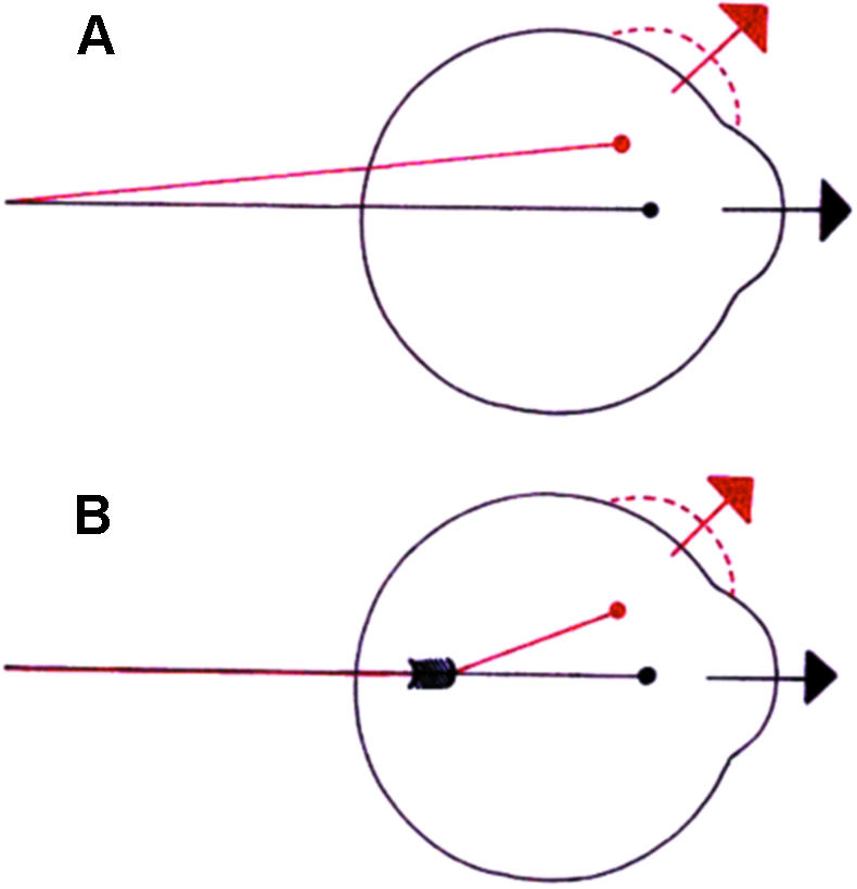
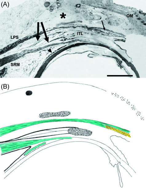
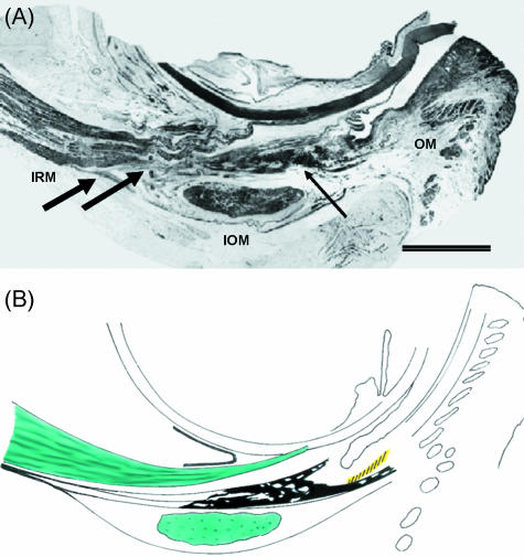
Recent studies have promoted the concept that rectus muscles pass through connective tissue pulleys located near the equator of the eye and act, in effect, as the muscle origins. Orbital muscle fibres (facing bone) terminate in pulleys, permitting adjustment of their position independent of the global fibres responsible for rotating the eye. The structure of pulleys (or muscle sleeves) and the passage taken by their muscle fibre insertions are unclear, and a detailed description is presented here together with a review of the active pulley hypothesis. Segments including the full width of single muscles were removed from the full orbital contents of dissection room cadavers and fresh perfusion-fixed rhesus and cynomolgus monkeys and prepared for light microscopy. Thin longitudinal sections were cut as facets from resin-embedded tissue blocks and montages assembled. Interrupted serial sections of selected regions of both species and ultrathin sections of monkey material were prepared for light and electron microscopy, respectively. Slender tendons leave the orbital surface of rectus muscles at intervals, aggregating and entering sleeves in humans and monkey; less frequently, tendons pass from the global surface to sleeves or insert directly in the posterior fascia bulbi. The orbital sides of sleeve rings are continuous with the fascial canopy of the globe and are 5-6 times as thick as the global sides; sleeve structure differs in the four recti. Medial rectus sleeves are the thickest, and contain smooth muscle, whereas little or none is present in the other rectus sleeves. Superior rectus sleeves are variable in structure and relatively insubstantial. A narrow interval separates muscles from the surrounding connective tissue equatorially in some preparations, consistent with a capacity to slide, but the tissues are contiguous in others, especially in monkey material. The structural organization of sleeves and their tendons, together with other presented factors, is inconsistent with a facility for the separate adjustment of sleeve position. The results favour the theory that sleeve tendons have just one role, to counter the viscoelastic resistance of global fascia - ocular and sleeve muscle fibres acting in unison. Whether the fragile sleeve structure can meet the physical demands of pulleys is questionable; but otherwise the veracity of the pulley hypothesis cannot be assessed from the structural relations of muscles and fascia bulbi reported.


Figures

Fig. 1
Fig. 1
Schematic representation showing (A) how the extraocular muscles were assumed to slip over the globe during eye rotation as described in the shortest path hypothesis by Boeder (1962) and (B) turning of tendon with no displacement as indicated by Miller (1989) using MRI scans.
Fig. 2
Fig. 2
(A) Photomontage of human medial rectus muscle (MRM). Fibres branch off from the orbital surface of the muscle (superior arrow) before merging into a compact collagenous mass of the muscle sleeve. The matrix diminishes and continues to the periosteum (P) as slender ligaments – the suspensory or check ligaments (arrowhead). Less substantial sleeve tendons issue from the global aspect of the muscle (inferior arrow) and reflect before inserting in the fascia bulbi. OM, orbicularis muscle. Scale bar = 5 mm. (B) Schematic representation of the medial rectus muscle as seen in A. Opposite the posterior pole the global and orbital sleeve thickness ratio is 1 : 6. Equatorially, the orbital sleeve thickens further to form a dense plate containing smooth muscle (yellow) and small lacunae filled with fat (white). The plate is attached to the orbital wall by fine ligaments (blue) looping behind the orbicularis muscle (stippled) to reach the periosteum.
Fig. 3
Fig. 3
Micrograph of the medial rectus muscle in monkey showing thin longitudinally orientated bundles of smooth muscle (arrow) embedded in the sleeve adjacent to the cartilage plate (CD). A small nictitans gland (N) is interposed between the plate and the conjunctiva (C). S, sclera. Scale bar = 400 µm.
Fig. 4
Fig. 4
(A) Photomontage of human lateral rectus muscle (LRM) showing fibres orbital and global fibres merging into the muscle sleeve (superior and inferior arrow, respectively). The distal portion of the orbital sleeve becomes thinner and slender extensions terminate opposite the fornix. Ligaments (L) attach the sleeve to the periosteum (P). Encapsulated lobules of lacrimal gland (LG) are interposed between the orbital sleeve and the orbicularis muscle (OM). Scale bar = 5 mm. (B) Schematic representation of the lateral muscle as seen in A. Opposite the posterior pole the orbital/global sheath thickness ratio is 5 : 1. The orbital sheath doubles in thickness at the equator. The sleeve is attached to periorbita by one or more strong ligaments (blue). There is little or no smooth muscle other than tarsal muscle (yellow).
Fig. 5
Fig. 5
(A) Photomontage of human superior rectus muscle showing strands of orbital tendons (thick arrows) advancing between the superior rectus muscle (SRM) and levator palpebrae superior muscle (LPS) before blending with the dense intermuscular transverse ligament (ITL). Small arrow indicates the position of the superior tarsal muscle. Asterisk, Whitnall's ligament; OM, orbicularis muscle; arrowhead, superior oblique. Scale bar = 5 mm. (B) Schematic representation of the superior rectus muscle as seen in A. Global sheath is delicate and the orbital sheath divides into several discrete bands that insert into the intermuscular transverse ligament. There are no direct attachments to the orbital wall. No smooth muscle is present other than superior tarsal muscle (yellow).
Fig. 6
Fig. 6
(A) Photomontage of human inferior rectus muscle (IRM) showing orbital tendons advancing between the inferior rectus and the inferior oblique muscle (IOM) while a minority of them pass under the inferior oblique (superior and inferior black arrows, respectively). The global tendon is thin and fibrous. Thin arrow indicates Lockwood's ligament. OM, orbicularis muscle. Scale bar = 5 mm. (B) Schematic representation of the inferior rectus muscle as seen in A. The global sheath is delicate and the thicker orbital sheath forms several discrete strands that insert into the suspensory ligament of Lockwood (triangular-shaped black structure). There are no direct attachments to the orbital wall. Smooth muscle is not present other than inferior tarsal muscle (yellow).

References

    1. Acheson GH. The topographical anatomy of the smooth muscle of the cat's nictitating membrane. Anat. Rec. 1938;71:297–311.
    1. Adachi B. Das Knorpelstück in der Plica semilunaris conjunctivae der Japaner. Z. Morph. Anthrop. 1906;9:325–326.
    1. Bartels P. Histolisch-anthropologische Untersuchungen der Plica semilunaris bei Herero und Hottentotten, sowie bei einigen Anthropoiden. Arch. Mikrosk. Anat. 1. Abt. 1911;78:29–564.
    1. Boeder P. Co-operative action of extraocular muscles. Br. J. Ophthalmol. 1962;46:397–403. - PMC - PubMed
    1. Clark RA, Miller JM, Demer JL. Three-dimensional location of human rectus pulleys by path inflections in secondary gaze positions. Invest. Ophthalmol. Vis. Sci. 2000;41:3787–3797. - PubMed

LinkOut - more resources