

Do malaria ookinete surface proteins P25 and P28 mediate parasite entry into mosquito midgut epithelial cells?
- PMID: 15733320
- PMCID: PMC555762
- DOI: 10.1186/1475-2875-4-15
Do malaria ookinete surface proteins P25 and P28 mediate parasite entry into mosquito midgut epithelial cells?
Abstract
Background: P25 and P28 are related ookinete surface proteins highly conserved throughout the Plasmodium genus that are under consideration as candidates for inclusion in transmission-blocking vaccines. Previous research using transgenic rodent malaria parasites lacking P25 and P28 has demonstrated that these proteins have multiple partially redundant functions during parasite infection of the mosquito vector, including an undefined role in ookinete traversal of the mosquito midgut epithelium, and it has been suggested that, unlike wild-type parasites, Dko P25/P28 parasites migrate across the midgut epithelium via an intercellular, rather than intracellular, route.
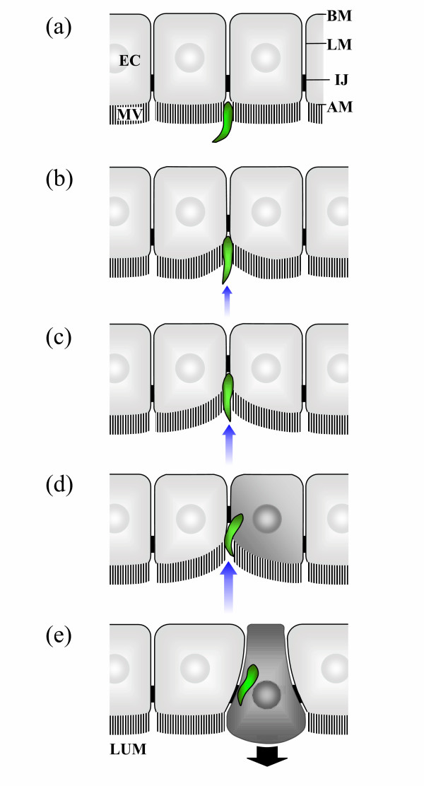
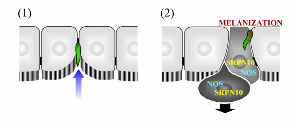
Presentation of the hypothesis: This paper presents an alternative interpretation for the previous observations of Dko P25/P28 parasites, based upon a recently published model of the route of ookinete invasion across the midgut epithelium. This model claims ookinete invasion is intracellular, with entry occurring through the lateral apical plasma membrane of midgut epithelial cells, and is associated with significant invagination of the midgut epithelium localised at the site of parasite penetration. Following this model, it is hypothesized that: (1) a sub-population of Dko P25/P28 ookinetes invaginate, but do not penetrate, the apical surface of the midgut epithelium and thus remain within the midgut lumen; and (2) another sub-population of Dko P25/P28 parasites successfully enters and migrates across the midgut epithelium via an intracellular route similar to wild-type parasites and subsequently develops into oocysts.
Testing the hypothesis: These hypotheses are tested by showing how they can account for previously published observations and incorporate them into a coherent and consistent explanatory framework. Based upon these hypotheses, several quantitative predictions are made, which can be experimentally tested, about the relationship between the densities of invading Dko P25/P28 ookinetes in different regions of the midgut epithelium and the number of oocyst stage parasites to which these mutant ookinetes give rise.
Implications of the hypothesis: The recently published model of ookinete invasion implies that Dko P25/P28 parasites are greatly, although not completely, impaired in their ability to enter the midgut epithelium. Therefore, P25 and/or P28 have a novel, previously unrecognized, function in mediating ookinete entry into midgut epithelial cells, suggesting that one mode of action of transmission-blocking antibodies to these ookinete surface proteins is to inhibit this function.
Figures
References
-
- Kaslow DC. Transmission-blocking vaccines. Chem Immunol. 2002;80:287–307. - PubMed
Publication types
MeSH terms
Substances
LinkOut - more resources
Full Text Sources
Medical
Research Materials
