Review
. 2005 Mar;58(3):225-36.
doi: 10.1136/jcp.2003.009506.

The links between axin and carcinogenesis

Affiliations
Review

The links between axin and carcinogenesis

S Salahshor et al. J Clin Pathol. 2005 Mar.

Erratum in

  • J Clin Pathol. 2005 Dec;58(12):1344

Abstract

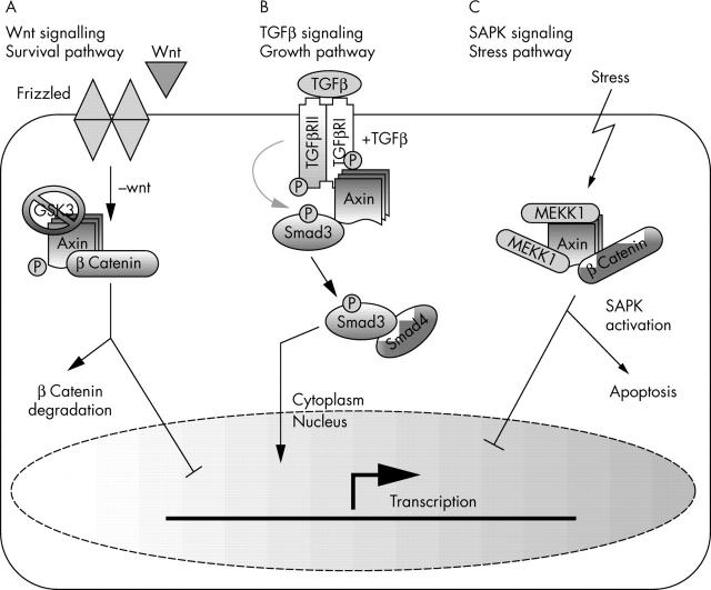
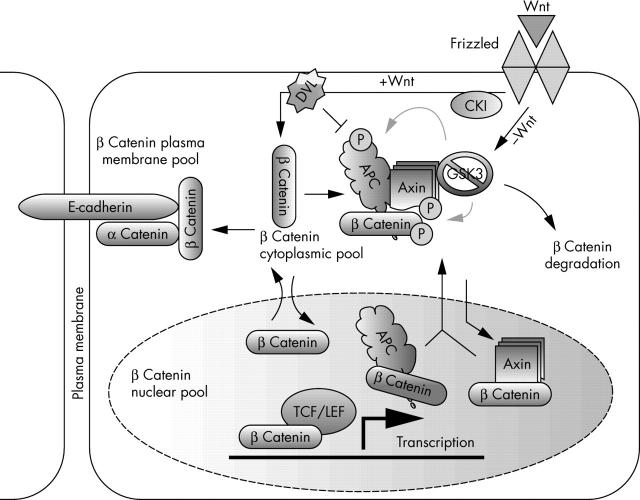
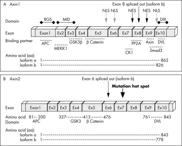
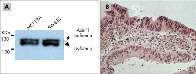
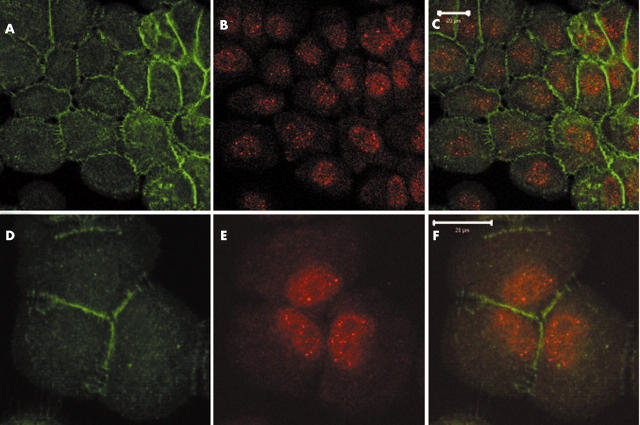
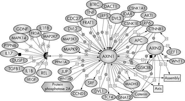
The products of the two mammalian Axin genes (Axin1 and its homologue Axin2) are essential for the degradation of beta catenin, a component of Wnt signalling that is frequently dysregulated in cancer cells. Axin is a multidomain scaffold protein that has many functions in biological signalling pathways. Overexpression of mutant [corrected] axin results in axis duplication in mouse embryos. Wnt signalling activity determines dorsal-ventral axis formation in vertebrates, implicating axin as a negative regulator of this signalling pathway. In addition, Wnts modulate pattern formation and the morphogenesis of most organs by influencing and controlling cell proliferation, motility, and fate. Defects in different components of the Wnt signalling pathway promote tumorigenesis and tumour progression. Recent biochemical studies of axins indicate that these molecules are the primary limiting components of this pathway. This review explores the intriguing connections between defects in axin function and human diseases.


Figures

Figure 1
Figure 1
At least three different signalling pathways are regulated by axin. (A) In the absence of Wnt ligands, axin stimulates β catenin phosphorylation and subsequent protease mediated degradation limits its transcriptional activity. (B) In the presence of transforming growth factor β (TGFβ) signals, axin stimulates Smad phosphorylation by TGFβ receptors (TGFβ receptors I and II). The activated Smads then translocate to the nucleus and activate transcription of downstream target genes. (C) In cells subjected to stress, axin binds to mitogen activated protein/extracellular regulated kinase kinase kinase 1 (MEKK1) and stimulates stress activated protein kinase (SAPK)/JNK (Jun N-terminal kinase) mediated apoptosis. GSK3, glycogen synthase kinase 3.
Figure 2
Figure 2
The role of axin in Wnt signalling. The Wnt signalling pathway plays an important role in the regulation of cellular proliferation, differentiation, motility, and morphogenesis. Axin serves as a scaffold protein that binds many of the proteins involved in this pathway. In the absence of Wnt ligands, the β catenin destruction complex, which is composed of adenomatous polyposis coli (APC), glycogen synthase kinase 3 (GSK3), and axin, is formed and leads to the phosphorylation and degradation of β catenin. In the presence of Wnt ligands, the formation of this complex is inhibited and the now stabilised β catenin is translocated into the nucleus and activates the transcription of downstream target genes. Axin is also implicated in shuttling β catenin out of the nucleus. However, β catenin can move in and out of the nucleus independently of axin. It is unclear whether axin participates in the regulation of all of the different pools of β catenin in the cell. Deregulation of many components of the Wnt pathway has been found in human cancer. DVL, Dishevelled; LEF, lymphoid enhancer binding factor; P, organic phosphate; TCF, T cell specific factor.
Figure 3
Figure 3
Genomic structure of Axin. (A) Axin1 is composed of 10 exons (encoding isoform a). Exon 8 is spliced out in isoform b. (B) Axin2 also consists of 10 exons that encode 843 amino acids (aa) (isoform a) or a 778 aa polypeptide (isoform b). Similar to Axin1, the binding partners of Axin2 are APC (aa 81–200), GSK3 (aa 372–413), β catenin (aa 414–476), and DVL (aa 761–843). So far, all mutations found in Axin2 are located in exon 7. APC, adenomatous polyposis coli; CKI, casein kinase I; DIX, Dishevelled and axin binding domain; DVL, Dishevelled; GSK3, glycogen synthase kinase 3; MEKK1, mitogen activated protein/extracellular regulated kinase kinase kinase 1; NES, nuclear export signal; NLS, nuclear localisation signal; PP2A, protein phosphatase.
Figure 4
Figure 4
Axin expression pattern and subcellular localisation in tumour cell lines and tumour tissue. (A) Western blot analysis of protein extracted from MCF12A and SW480 cells confirmed the existence of two axin1 isoforms (a and b). (B) The subcellular localisation of axin1 was examined in a sporadic colorectal cancer case and axin1 was found to have a predominantly nuclear localisation.
Figure 5
Figure 5
Subcellular localisation of axin1 in MCF12A cells. Based on the known function of axin1, it is expected to be mainly localised in the cytoplasm. Confocal sections of MCF12A cells stained with (A and D) anti-axin1 and (B and E) anti-β catenin antibodies (C and F merged) show both nuclear and cytoplasmic axin1 staining, with a pronounced nuclear localisation, whereas β catenin is located mainly at the cell membrane (scale bars, 20 μm). The following antibodies were used: polyclonal anti-axin1 (1/500 dilution; Zymed, South San Francisco, California, USA), monoclonal anti-β catenin antibody (1/500 dilution; Signal Transduction Laboratories; Lexington, Kentucky, USA), secondary monoclonal and polyclonal antibodies (1/1000 dilution; Molecular Probes, Eugene, Oregon, USA). Other studies show a more cytoplasmic localisation for axin1. However, the subcellular localisation of axin1 appears to be cell type dependent.
Figure 6
Figure 6
Axin regulators and binding partners. To bring all the relevant information about axin together, we have used PathwayAssist software (Stratagene) to generate a network of molecules that are either positively (+) or negatively (−) regulated by axin. Each interaction is annotated with the source references and contains hyperlinks to the source literature on PubMed. The biological data have been extracted from PubMed. The references that support the interaction or regulation for each molecule can be viewed by holding the mouse on the plus or minus sign. This pathway is an online navigation tool (http://kinase.uhnres.utoronto.ca/Sima/axins_pathway/).

Similar articles

Cited by

References

    1. Zeng L, Fagotto F, Zhang T, et al. The mouse Fused locus encodes axin, an inhibitor of the Wnt signalling pathway that regulates embryonic axis formation. Cell 1997;90:181–92. - PubMed
    1. Satoh S, Daigo Y, Furukawa Y, et al. AXIN1 mutations in hepatocellular carcinomas, and growth suppression in cancer cells by virus-mediated transfer of AXIN1. Nat Genet 2000;24:245–50. - PubMed
    1. Akiyama T . Wnt/beta-catenin signaling. Cytokine Growth Factor Rev 2000;11:273–82. - PubMed
    1. Wodarz A, Nusse R. Mechanisms of Wnt signaling in development. Annu Rev Cell Dev Biol 1998;14:59–88. - PubMed
    1. Bienz M, Clevers H. Linking colorectal cancer to Wnt signaling. Cell 2000;103:311–20. - PubMed

Publication types

MeSH terms