

Phenotypic analysis of bovine chondrocytes cultured in 3D collagen sponges: effect of serum substitutes
- PMID: 15735900
- PMCID: PMC1242111
- DOI: 10.1007/s10561-005-5810-0
Phenotypic analysis of bovine chondrocytes cultured in 3D collagen sponges: effect of serum substitutes
Abstract
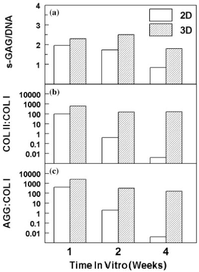
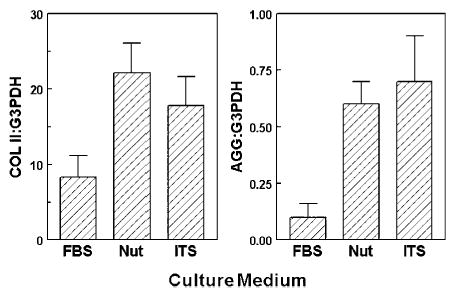
Repair of damaged cartilage usually requires replacement tissue or substitute material. Tissue engineering is a promising means to produce replacement cartilage from autologous or allogeneic cell sources. Scaffolds provide a three-dimensional (3D) structure that is essential for chondrocyte function and synthesis of cartilage-specific matrix proteins (collagen type II, aggrecan) and sulfated proteoglycans. In this study, we assessed porous, 3D collagen sponges for in vitro engineering of cartilage in both standard and serum-free culture conditions. Bovine articular chondrocytes (bACs) cultured in 3D sponges accumulated and maintained cartilage matrix over 4 weeks, as assessed by quantitative measures of matrix content, synthesis, and gene expression. Chondrogenesis by bACs cultured with Nutridoma as a serum replacement was equivalent or better than control cultures in serum. In contrast, chondrogenesis in insulin-transferrin-selenium (ITS(+3)) serum replacement cultures was poor, apparently due to decreased cell survival. These data indicate that porous 3D collagen sponges maintain chondrocyte viability, shape, and synthetic activity by providing an environment favorable for high-density chondrogenesis. With quantitative assays for cartilage-specific gene expression and biochemical measures of chondrogenesis in these studies, we conclude that the collagen sponges have potential as a scaffold for cartilage tissue engineering.
Figures
References
-
- Benya PD, Padilla SR, Nimni ME. Independent regulation of collagen types by chondrocytes during the loss of differentiated function in culture. Cell. 1978;15:1313–1321. - PubMed
-
- Benya PD, Shaffer JD. Dedifferentiated chondrocytes reexpress the differentiated collagen phenotype when cultured in agarose gels. Cell. 1982;30:215–224. - PubMed
-
- Boyan BD, Hummert TW, Dean DD, Schwartz Z. Role of material surfaces in regulating bone and cartilage cell response. Biomaterials. 1996;17:137–146. - PubMed
-
- Brittberg M, Lindahl A, Nilsson A, Ohlsson C, Isaksson O, Peterson L. Treatment of deep cartilage defects in the knee with autologous chondrocyte transplantation. N Engl J Med. 1994;331:889–895. - PubMed
-
- Brittberg M, Peterson L, Sjogren-Jansson E, Tallheden T, Lindahl A. Articular cartilage engineering with autologous chondrocyte transplantation. A review of recent developments. J Bone Joint Surg Am. 2003;85A(Suppl 3):109–115. - PubMed
Publication types
MeSH terms
Substances
Grants and funding
LinkOut - more resources
Full Text Sources
