. 2005 Mar;6(1):48-62.
doi: 10.1007/s10162-004-5009-2. Epub 2005 Apr 22.

Disruption of lateral olivocochlear neurons via a dopaminergic neurotoxin depresses sound-evoked auditory nerve activity

Affiliations

Disruption of lateral olivocochlear neurons via a dopaminergic neurotoxin depresses sound-evoked auditory nerve activity

Colleen G Le Prell et al. J Assoc Res Otolaryngol. 2005 Mar.

Abstract

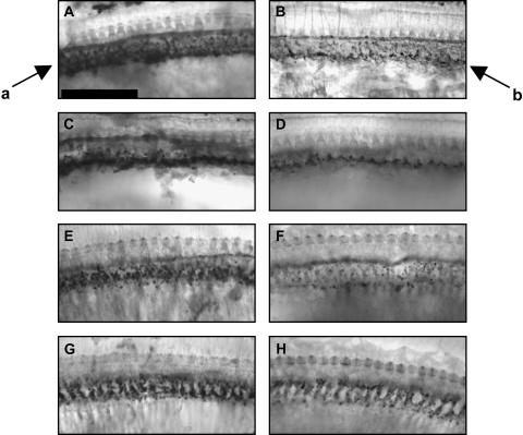
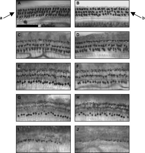
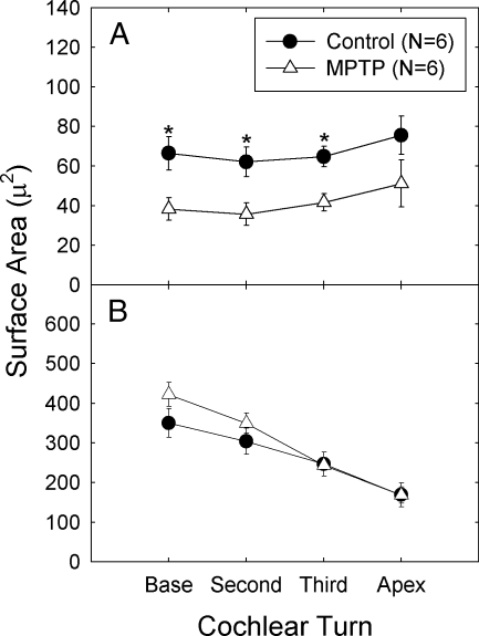
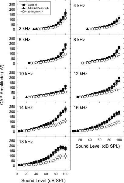
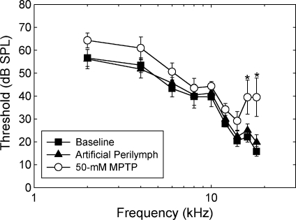
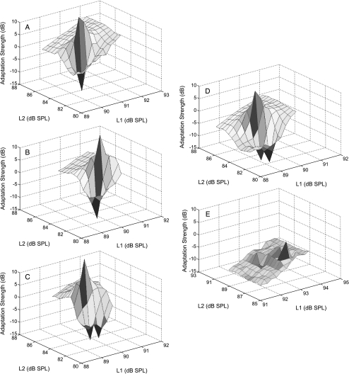
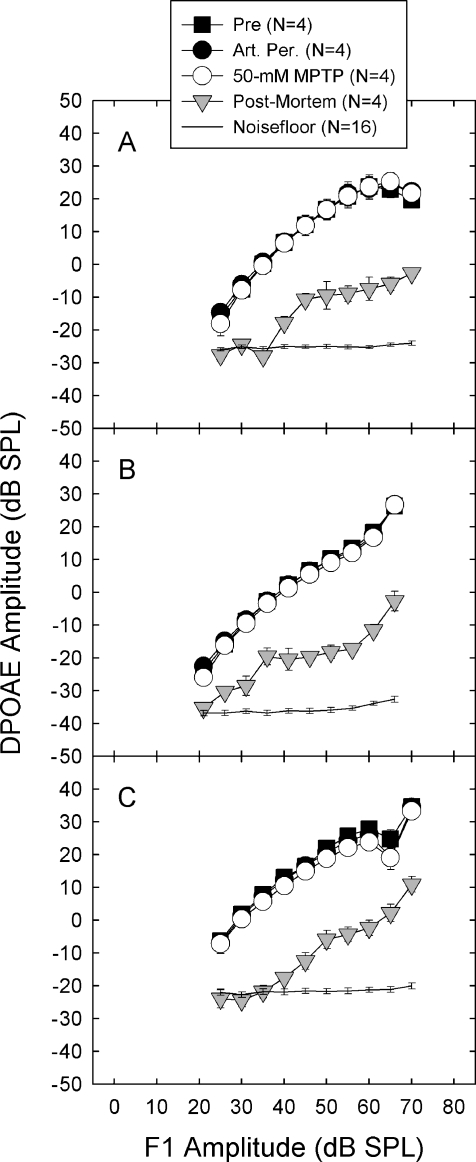
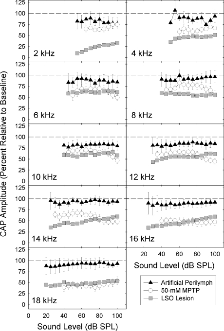
We applied the dopaminergic (DA) neurotoxin 1-methyl-4-phenyl-1,2,3,6-tetrahydropyridine (MPTP) to the guinea pig cochlear perilymph. Immunolabeling of lateral olivocochlear (LOC) neurons using antibodies against synaptophysin was reduced after the MPTP treatment. In contrast, labeling of the medial olivocochlear innervation remained intact. As after brainstem lesions of the lateral superior olive (LSO), the site of origin of the LOC neurons, the main effect of disrupting LOC innervation of the cochlea via MPTP was a depression of the amplitude of the compound action potential (CAP). CAP amplitude depression was similar to that produced by LSO lesions. Latency of the N1 component of the CAP, and distortion product otoacoustic emission amplitude and adaptation were unchanged by the MPTP treatment. This technique for selectively lesioning descending LOC efferents provides a new opportunity for examining LOC modulation of afferent activity and behavioral measures of perception.


Figures

Fig. 1
Fig. 1
Cochlear tissues had less dense immunolabeling of lateral olivocochlear (LOC) efferents after MPTP. Tissues were immunolabeled with antisynaptophysin, and are from base (A, B), second (C, D) and third (E, F) turns, and apex (G, H). Control tissues are depicted in the left panels (A, C, E, G). MPTP-treated tissues are depicted in the right panels (B, D, F, H). Arrows indicate normal LOC immunolabeling below the inner hair cells in the region of the inner spiral bundle (arrow a) and sparse LOC immunolabeling after MPTP treatment (arrow b). Scale bar = 50 μm.
Fig. 2
Fig. 2
Medial olivocochlear (MOC) immunolabeling was not affected by MPTP. Tissues were immunolabeled with antisynaptophysin, and are from base (A, B), second (C, D) and third (E, F) turns, lower apex (G, H) and upper apex (I, J). Control tissues are depicted in the left panels (A, C, E, G, H). MPTP-treated tissues are depicted in the right panels (B, D, F, H, J). Arrows indicate normal MOC immunolabeling below the outer hair cells (arrow a), and unchanged immunolabeling of MOC puncta after MPTP treatment (arrow b). Scale bar = 50 μm.
Fig. 3
Fig. 3
A. Surface area of synaptophysin immunolabeling of the lateral olivocochlear (LOC) neurons was reduced in MPTP-treated cochlear tissues. B. Surface area of medial olivocochlear (MOC) neurons was not reliably changed. Asterisks indicate statistically reliable differences between control and MPTP-treated ears. The amount of tissue from apical turns was not sufficient for the conduct of statistical comparisons, and all hook region tissues were excluded from analysis as the majority of hook region tissues from both the control and MPTP groups were damaged during dissection. For all other tissues, labeled area was measured in three to six regions of each cochlear turn for each animal. Labeling was first averaged within each animal; average labeling across animals is depicted.
Fig. 4
Fig. 4
Amplitude of the compound action potential (CAP) amplitude, defined as the amplitude of the N1–P1 component, was depressed after MPTP treatment. CAP amplitude was determined from 0- to 100-dB SPL at frequencies extending from 2 to 18 kHz. Mean CAP amplitude (±SE) is depicted immediately after cementing the recording electrode in place, 30 min after applying artificial perilymph to the round window membrane, and 30 min after applying 50 mM MPTP to the round window membrane.
Fig. 5
Fig. 5
Threshold (±SE) of the compound action potential (CAP) was elevated by MPTP treatment only at the highest frequencies tested. Asterisks indicate statistically reliable differences between control and MPTP-treated ears. CAP threshold was determined immediately after cementing the recording electrode in place, 30 min after applying artificial perilymph to the round window membrane, and 30 min after applying 50 mM MPTP to the round window membrane.
Fig. 6
Fig. 6
Onset adaptation of distortion product otoacoustic emissions (DPOAEs) was not reliably changed by MPTP treatment. DPOAEs show a rapid level-dependant adaptation shortly after signal onset when the medial olivocochlear (MOC) pathway is intact. DPOAE adaptation is depicted 30 min after sequential applications of artificial perilymph (panels A, B, C), 30 min after applying 50 mM MPTP to the round window membrane (D), and after i.v. strychnine (0.15 mg/kg, see panel E).
Fig. 7
Fig. 7
Steady-state amplitude of distortion product otoacoustic emissions (DPOAE) was unchanged by MPTP treatment. Steady-state DPOAE amplitude is a measure of the integrity of the outer hair cell population. DPOAE steady-state amplitude is depicted after opening the bulla (“Pre”), 30 min after applying artificial perilymph to the round window membrane, and 30 min after applying 50 mM MPTP to the round window membrane. The levels of F1 and F2 were systematically varied such that F2 was always 10 dB quieter than F1. DPOAE frequencies were 5.8 kHz (A), 8.7 kHz (B), and 11.7 kHz (C).
Fig. 8
Fig. 8
Depression of the amplitude of the compound action potential (CAP) was generally equivalent in animals treated with intracochlear MPTP and animals in which the lateral superior olive (LSO) was lesioned. Amplitude of the CAP 30 min after applying artificial perilymph or 50 mM MPTP to the round window membrane was normalized to pretreatment baseline. We also normalized CAP amplitude assessed 1 week after lesioning the LSO to baseline measures from animals in which the LSO was intact (original data are presented in Le Prell et al. 2003b). Dashed lines indicate 100% of baseline; decreasing values indicate CAP was depressed post-treatment.

Similar articles

Cited by

References

    1. Abou-Madi L, Pontarotti P, Tramu G, Cupo A, Eybalin M. Coexistence of putative neuroactive substances in lateral olivocochlear neurons of rat and guinea pig. Hear. Res. 1987;30:135–146. - PubMed
    1. Adams JC, Mroz EA, Sewell WF. A possible neurotransmitter role for CGRP in a hair-cell sensory organ. Brain Res. 1987;419:347–351. - PubMed
    1. Altschuler RA, Parakkal MH, Fex J. Localization of enkephalin-like immunoreactivity in acetylcholinesterase-positive cells in the guinea-pig lateral superior olivary complex that project to the cochlea. Neuroscience. 1983;9:621–630. - PubMed
    1. Altschuler RA, Fex J, Parakkal MH, Eckenstein F. Colocalization of enkephalin-like and choline acetyltransferase-like immunoreactivities in olivocochlear neurons of the guinea pig. J. Histochem. Cytochem. 1984;32:839–843. - PubMed
    1. Altschuler RA, Hoffman DW, Reeks KA, Fex J. Localization of dynorphin B-like and alpha-neoendorphin-like immunoreactivities in the guinea pig organ of Corti. Hear. Res. 1985;17:249–258. - PubMed

Publication types

MeSH terms