

Reverse engineering the L-type Ca2+ channel alpha1c subunit in adult cardiac myocytes using novel adenoviral vectors
- PMID: 15737650
- PMCID: PMC2751644
- DOI: 10.1016/j.bbrc.2005.02.015
Reverse engineering the L-type Ca2+ channel alpha1c subunit in adult cardiac myocytes using novel adenoviral vectors
Abstract
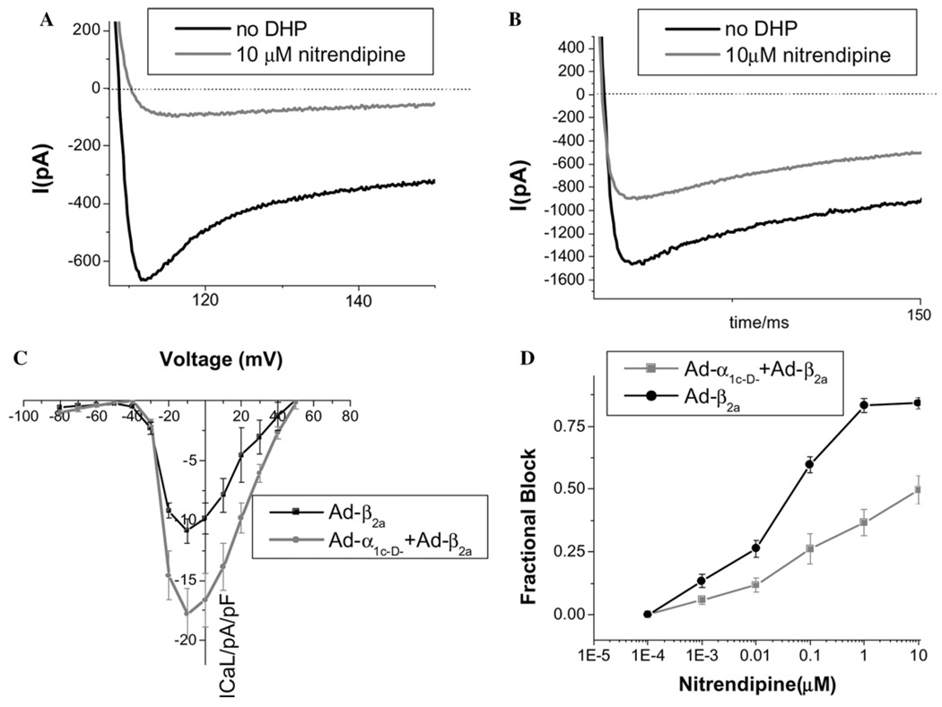
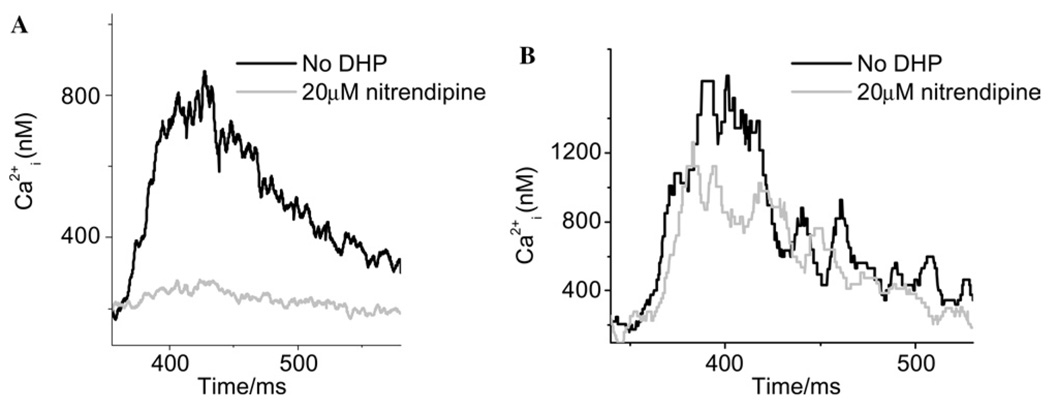
The alpha(1c) subunit of the cardiac L-type Ca(2+) channel, which contains the channel pore, voltage- and Ca(2+)-dependent gating structures, and drug binding sites, has been well studied in heterologous expression systems, but many aspects of L-type Ca(2+) channel behavior in intact cardiomyocytes remain poorly characterized. Here, we develop adenoviral constructs with E1, E3 and fiber gene deletions, to allow incorporation of full-length alpha(1c) gene cassettes into the adenovirus backbone. Wild-type (alpha(1c-wt)) and mutant (alpha(1c-D-)) Ca(2+) channel adenoviruses were constructed. The alpha(1c-D-) contained four point substitutions at amino acid residues known to be critical for dihydropyridine binding. Both alpha(1c-wt) and alpha(1c-D-) expressed robustly in A549 cells (peak L-type Ca(2+) current (I(CaL)) at 0 mV: alpha(1c-wt) -9.94+/-1.00pA/pF, n=9; alpha(1c-D-) -10.30pA/pF, n=12). I(CaL) carried by alpha(1c-D-) was markedly less sensitive to nitrendipine (IC(50) 17.1 microM) than alpha(1c-wt) (IC(50) 88 nM); a feature exploited to discriminate between engineered and native currents in transduced guinea-pig myocytes. 10 microM nitrendipine blocked only 51+/-5% (n=9) of I(CaL) in alpha(1c-D-)-expressing myocytes, in comparison to 86+/-8% (n=9) of I(CaL) in control myocytes. Moreover, in 20 microM nitrendipine, calcium transients could still be evoked in alpha(1c-D-)-transduced cells, but were largely blocked in control myocytes, indicating that the engineered channels were coupled to sarcoplasmic reticular Ca(2+) release. These alpha(1c) adenoviruses provide an unprecedented tool for structure-function studies of cardiac excitation-contraction coupling and L-type Ca(2+) channel regulation in the native myocyte background.
Figures


Similar articles
-
Cardiac-type EC-coupling in dysgenic myotubes restored with Ca2+ channel subunit isoforms alpha1C and alpha1D does not correlate with current density.Biophys J. 2003 Jun;84(6):3816-28. doi: 10.1016/S0006-3495(03)75109-1. Biophys J. 2003. PMID: 12770887 Free PMC article.
-
Triadin overexpression stimulates excitation-contraction coupling and increases predisposition to cellular arrhythmia in cardiac myocytes.Circ Res. 2005 Apr 1;96(6):651-8. doi: 10.1161/01.RES.0000160609.98948.25. Epub 2005 Feb 24. Circ Res. 2005. PMID: 15731460
-
New Determinant for the CaVbeta2 subunit modulation of the CaV1.2 calcium channel.J Biol Chem. 2008 Jun 6;283(23):15577-88. doi: 10.1074/jbc.M802035200. Epub 2008 Apr 14. J Biol Chem. 2008. PMID: 18411278 Free PMC article.
-
Ca2+ currents in cardiac myocytes: Old story, new insights.Prog Biophys Mol Biol. 2006 May-Jun;91(1-2):1-82. doi: 10.1016/j.pbiomolbio.2005.01.001. Epub 2005 Feb 25. Prog Biophys Mol Biol. 2006. PMID: 16503439 Review.
-
L-type Ca2+ channels in the heart: structure and regulation.Medicina (Kaunas). 2008;44(7):491-9. Medicina (Kaunas). 2008. PMID: 18695345 Review. English, Lithuanian.
Cited by
-
The PDZ motif of the α1C subunit is not required for surface trafficking and adrenergic modulation of CaV1.2 channel in the heart.J Biol Chem. 2015 Jan 23;290(4):2166-74. doi: 10.1074/jbc.M114.602508. Epub 2014 Dec 11. J Biol Chem. 2015. PMID: 25505241 Free PMC article.
-
Sympathetic Nervous System Regulation of Cardiac Calcium Channels.Handb Exp Pharmacol. 2023;279:59-82. doi: 10.1007/164_2022_632. Handb Exp Pharmacol. 2023. PMID: 36592229
-
Functional expression of transgenic 1sDHPR channels in adult mammalian skeletal muscle fibres.J Physiol. 2011 Mar 15;589(Pt 6):1421-42. doi: 10.1113/jphysiol.2010.202804. Epub 2011 Jan 24. J Physiol. 2011. PMID: 21262876 Free PMC article.
-
Manipulating L-type calcium channels in cardiomyocytes using split-intein protein transsplicing.Proc Natl Acad Sci U S A. 2013 Sep 17;110(38):15461-6. doi: 10.1073/pnas.1308161110. Epub 2013 Sep 3. Proc Natl Acad Sci U S A. 2013. PMID: 24003157 Free PMC article.
-
Beta-adrenergic stimulation of L-type Ca2+ channels in cardiac myocytes requires the distal carboxyl terminus of alpha1C but not serine 1928.Circ Res. 2006 Feb 3;98(2):e11-8. doi: 10.1161/01.RES.0000202692.23001.e2. Epub 2006 Jan 5. Circ Res. 2006. PMID: 16397147 Free PMC article.
References
-
- Kamp TJ, Hell JW. Circ. Res. 2000;87:1095–1102. - PubMed
-
- Hille B. Ionic Channels of Excitable Membranes. Sunderland, MA: Sinauer Associates, Inc; 1992.
-
- Bers DM. Excitation-Contraction Coupling and Cardiac Contractile Force. second ed. Dordrecht, Netherlands: Kluwer; 2001.
-
- Striessnig J, Grabner M, Mitterdorfer J, Hering S, Sinnegger MJ, Glossmann H. Trends Pharmacol. Sci. 1998;19:108–115. - PubMed
Publication types
MeSH terms
Substances
Grants and funding
LinkOut - more resources
Full Text Sources
Miscellaneous
