

Reverse engineering the L-type Ca2+ channel alpha1c subunit in adult cardiac myocytes using novel adenoviral vectors
- PMID: 15737650
- PMCID: PMC2751644
- DOI: 10.1016/j.bbrc.2005.02.015
Reverse engineering the L-type Ca2+ channel alpha1c subunit in adult cardiac myocytes using novel adenoviral vectors
Abstract
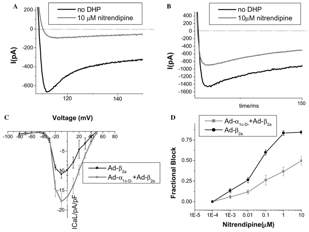
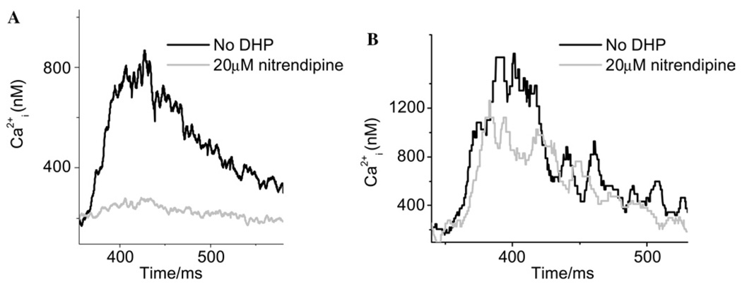
The alpha(1c) subunit of the cardiac L-type Ca(2+) channel, which contains the channel pore, voltage- and Ca(2+)-dependent gating structures, and drug binding sites, has been well studied in heterologous expression systems, but many aspects of L-type Ca(2+) channel behavior in intact cardiomyocytes remain poorly characterized. Here, we develop adenoviral constructs with E1, E3 and fiber gene deletions, to allow incorporation of full-length alpha(1c) gene cassettes into the adenovirus backbone. Wild-type (alpha(1c-wt)) and mutant (alpha(1c-D-)) Ca(2+) channel adenoviruses were constructed. The alpha(1c-D-) contained four point substitutions at amino acid residues known to be critical for dihydropyridine binding. Both alpha(1c-wt) and alpha(1c-D-) expressed robustly in A549 cells (peak L-type Ca(2+) current (I(CaL)) at 0 mV: alpha(1c-wt) -9.94+/-1.00pA/pF, n=9; alpha(1c-D-) -10.30pA/pF, n=12). I(CaL) carried by alpha(1c-D-) was markedly less sensitive to nitrendipine (IC(50) 17.1 microM) than alpha(1c-wt) (IC(50) 88 nM); a feature exploited to discriminate between engineered and native currents in transduced guinea-pig myocytes. 10 microM nitrendipine blocked only 51+/-5% (n=9) of I(CaL) in alpha(1c-D-)-expressing myocytes, in comparison to 86+/-8% (n=9) of I(CaL) in control myocytes. Moreover, in 20 microM nitrendipine, calcium transients could still be evoked in alpha(1c-D-)-transduced cells, but were largely blocked in control myocytes, indicating that the engineered channels were coupled to sarcoplasmic reticular Ca(2+) release. These alpha(1c) adenoviruses provide an unprecedented tool for structure-function studies of cardiac excitation-contraction coupling and L-type Ca(2+) channel regulation in the native myocyte background.
Figures


References
-
- Kamp TJ, Hell JW. Circ. Res. 2000;87:1095–1102. - PubMed
-
- Hille B. Ionic Channels of Excitable Membranes. Sunderland, MA: Sinauer Associates, Inc; 1992.
-
- Bers DM. Excitation-Contraction Coupling and Cardiac Contractile Force. second ed. Dordrecht, Netherlands: Kluwer; 2001.
-
- Striessnig J, Grabner M, Mitterdorfer J, Hering S, Sinnegger MJ, Glossmann H. Trends Pharmacol. Sci. 1998;19:108–115. - PubMed
Publication types
MeSH terms
Substances
Grants and funding
LinkOut - more resources
Full Text Sources
Miscellaneous
