. 2005 Mar 7;201(5):793-804.
doi: 10.1084/jem.20042034. Epub 2005 Feb 28.

Phosphorylation of histone deacetylase 7 by protein kinase D mediates T cell receptor-induced Nur77 expression and apoptosis

Affiliations

Phosphorylation of histone deacetylase 7 by protein kinase D mediates T cell receptor-induced Nur77 expression and apoptosis

Franck Dequiedt et al. J Exp Med. .

Abstract

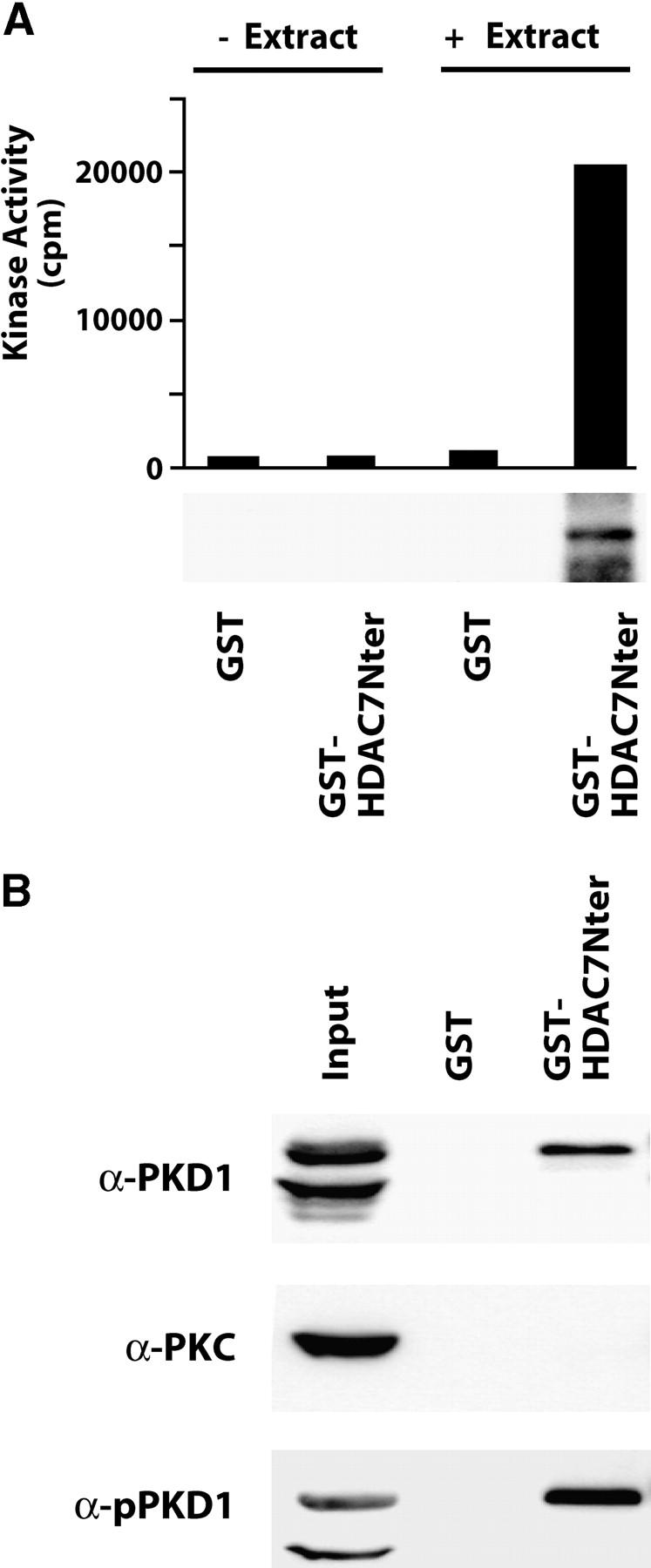
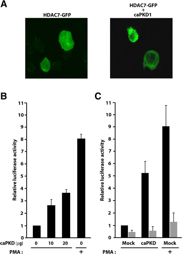
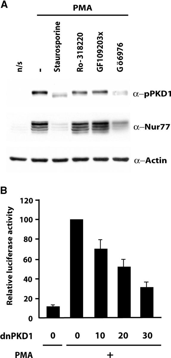
The molecular basis of thymocyte negative selection, a crucial mechanism in establishing central tolerance, is not yet resolved. Histone deacetylases (HDACs) have emerged as key transcriptional regulators in several major developmental programs. Recently, we showed that the class IIa member, HDAC7, regulates negative selection by repressing expression of Nur77, an orphan nuclear receptor involved in antigen-induced apoptosis of thymocytes. Engagement of the T cell receptor (TCR) alleviates this repression through phosphorylation-dependent nuclear exclusion of HDAC7. However, the identity of the TCR-activated kinase that phosphorylates and inactivates HDAC7 was still unknown. Here, we demonstrate that TCR-induced nuclear export of HDAC7 and Nur77 expression is mediated by activation of protein kinase D (PKD). Indeed, active PKD stimulates HDAC7 nuclear export and Nur77 expression. In contrast, inhibition of PKD prevents TCR-mediated nuclear exclusion of HDAC7 and associated Nur77 activation. Furthermore, we show that HDAC7 is an interaction partner and a substrate for PKD. We identify four serine residues in the NH(2) terminus of HDAC7 as targets for PKD. More importantly, a mutant of HDAC7 specifically deficient in phosphorylation by PKD, inhibits TCR-mediated apoptosis of T cell hybridomas. These findings indicate that PKD is likely to play a key role in the signaling pathways controlling negative selection.


Figures

Figure 1.
Figure 1.
TCR-induced nuclear export of HDAC7 is mediated by a PMA-stimulated and Gö6976-sensitive kinase. (A) Polyclonal DO11.10 cells stably expressing HDAC7-GFP were untreated or treated with anti-CD3 antibodies, PMA and ionomycin, ionomycin, or PMA. Subcellular localization of GFP-HDAC7 was monitored by confocal microscopy after 2 h of treatment. Bar histograms represent the mean percentages of cells showing nuclear exclusion of HDAC7-GFP in each condition. (B) Polyclonal DO11.10 cells stably expressing HDAC7-GFP were treated with PMA with or without previous incubation with the indicated inhibitors. After 2 h of PMA treatment, localization of HDAC7 was then examined using confocal microscopy. Bar histograms represent the mean percentages of cells showing nuclear exclusion of HDAC7-GFP in each condition.
Figure 2.
Figure 2.
PKD is expressed and activated by TCR in thymocytes. (A) DO11.10 cells were left unstimulated (n/s) or stimulated for 15 min with anti-CD3 antibodies, PMA and ionomycin, PMA alone, or ionomycin alone. Total protein extracts were analyzed by Western blotting with an antibody specific for phosphorylated serine 916 of PKD1 (pPKD1, which recognizes active PKD1). As a control, the same membrane was stripped and probed with an antiserum against total PKD1. (B) DO11.10 cells were cultured in the presence of 10 ng/ml PMA, and activation of PKD1 was examined at 0, 2, 5, 15, 30, 120, and 240 min by Western blot using the phosphorylated serine 916–specific antibody. The same blot was probed with an antibody for total PKD1 as control.
Figure 3.
Figure 3.
PKD associates with the NH2 terminus of HDAC7. (A) The NH2 terminus of HDAC7 (aa 1–490) was expressed as a GST fusion protein and immobilized on glutathione agarose beads. GST protein alone was used as control. Before (− Extract) and after (+ Extract) incubation of equal amounts of the fusion proteins with total cellular extracts from unstimulated, actively growing DO11.10 cells, associated kinase activity was revealed by IVK. Total incorporated radioactivity was quantified by scintillation counting. Results representative of three independent experiments are shown as a histogram (top). In parallel, IVK assays were analyzed by SDS-PAGE and autoradiography (bottom). (B) Pull-downs were analyzed for the presence of endogenous PKD1, PKC family members (PKC), and activated PKD1 (pPKD1) by Western blot.
Figure 4.
Figure 4.
PKD1 phosphorylates S155, S181, S321, and S449 of HDAC7 in vitro. (A) The C- (aa 490–915) and N- (aa 1–490) terminal domains of HDAC7 were produced as GST fusion proteins (GST-HDAC7Cter and GST-HDAC7Nter, respectively) and immobilized on glutathione beads. Equal amounts of purified recombinant proteins were used in IVK assays with constitutively active recombinant PKD1. IVK reactions were analyzed by SDS-PAGE and Coomassie staining (left) before autoradiography (right). (B) Sequences around the four identified serines in HDAC7 match with the canonical PKD recognition motif. (C) GST fusion proteins corresponding to the sequences surrounding the four putative PKD phosphorylation sites in HDAC7 were used as substrates in independent IVK assays with constitutively active recombinant PKD1. Mutant fusion proteins harboring a serine to alanine substitution were used as controls. Reactions were resolved by SDS-PAGE and gel was stained with Coomassie, dried, and analyzed by autoradiography. The arrow indicates the radioactive band corresponding to autophosphorylated PKD1 (rPKD1).
Figure 5.
Figure 5.
PKD activation is associated with HDAC7 nuclear export and derepression of the Nur77 promoter. (A) DO11.10 cells were transfected with an expression vector encoding GFP-HDAC7 in the absence or presence of a plasmid for activated PKD1. Subcellular localization of HDAC7 was examined by confocal microscopy. (B) Increasing amounts of an expression vector for activated PKD1 were cotransfected with a luciferase reporter plasmid driven by the Nur77 promoter (pNur77-Luc) into DO11.10 cells. The activity of the Nur77 promoter is shown relative to its basal activity, measured in the absence of activated PKD1 using dual luciferase assay. Luciferase activities were measured in untreated (−) cells or cells treated with PMA for 4 h (+). Results are from at least three independent experiments each performed in triplicate. (C) DO11.10 cells were cotransfected with Nur77-responsive reporter constructs pNBREwt-Luc (black bars) or pNBREmut-Luc (gray bars) along with an expression vector for active PKD1. Luciferase activities are presented relative to the basal luciferase activity of pNBREwt-Luc, measured in the absence of active PKD1 using the dual luciferase assay. Luciferase activities were measured in untreated (−) cells or cells treated with PMA for 4 h (+). Results are from at least three independent experiments each performed in triplicate.
Figure 6.
Figure 6.
Repression of PKD is associated with reduced Nur77 promoter activity. (A) DO11.10 cells were left unstimulated (n/s) or treated with PMA with or without (−) previous incubation with the indicated inhibitors. After 2 h of PMA treatment, total cellular extracts were prepared and analyzed by Western blotting with antisera for activated PKD1 (pPKD1), Nur77, and actin. (B) DO11.10 cells were transfected with pNur77-Luc and increasing amounts of an expression vector coding for a dominant negative mutant of PKD1. 40 h after transfection, cells were left unstimulated (−) or activated with PMA for 4 h (+). The activity of the Nur77 promoter is shown relative to its activity after PMA treatment in the absence of dominant negative PKD1 measured with the dual luciferase assay. Results are from three independent experiments each performed in triplicate.
Figure 7.
Figure 7.
PKD regulates Nur77 expression in response to TCR stimulation. (A) GST fusion proteins corresponding to the sequences surrounding the four putative PKD phosphorylation sites in HDAC7 were used independently as substrates in IVK assays with constitutively active recombinant PKD1. Corresponding mutant fusion proteins, harboring a leucine to alanine substitution at position −5 relative to the phosphorylated serine, were tested in parallel. IVK reactions were resolved by SDS-PAGE. The gel was stained with Coomassie, dried, and analyzed by autoradiography. The arrow indicates the radioactive band corresponding to autophosphorylated PKD1. (B) Polyclonal DO11.10 cells stably expressing wild-type HDAC7 (HDAC7wt) or the L150/176/316/444A HDAC7 mutant (HDAC7ΔL), both fused to GFP, were treated with PMA. Subcellular localization of fluorescent proteins was monitored by confocal microscopy after 2 h of treatment. Bar histograms represent the mean percentages of cells showing nuclear exclusion. (C) Polyclonal DO11.10 cell lines, stably expressing FLAG-tagged versions of HDAC7 (wt) and the HDAC7 mutant (HDAC7ΔL) or the empty vector (Empty), were left unstimulated (−) or stimulated with PMA (+) for 1. 5 h. Total cellular extracts were prepared and analyzed by Western blotting with antisera specific for FLAG, Nur77, and actin. (D) DO11.10 cells were transfected with the pNur77-Luc reporter construct and the same quantity of empty vector (Mock), wild-type HDAC7 (WT), or the ΔL mutant (ΔL). Luciferase activity was measured in cells treated with PMA (left, +) or ionomycin (right, +) and untreated cells (−). The activity of the Nur77 promoter is expressed relative to its activity in the mock-transfected cells after PMA (left) or ionomycin (right) treatment as measured by dual luciferase assay. Results are from three independent experiments each performed in triplicate.
Figure 8.
Figure 8.
PKD regulates TCR-mediated apoptosis. (A) Polyclonal DO11.10 cells lines, stably expressing wild-type HDAC7 (WT), HDAC7ΔL (ΔL), or the empty vector (Mock) were left untreated or treated with PMA/ionomycin, anti-CD3 antibodies, or cycloheximide (CHX) for 20 h. Apoptotic cells were detected by PE–annexin V staining and flow cytometry. Flow histograms illustrate the percentages of apoptotic cells detected in one representative experiment. (B) Mean apoptotic rates from nine independent experiments (as described in A) performed from three independently established set of polyclonal cell lines are shown. Results are expressed relative to the apoptotic rate observed in the mock-transduced cell line and are shown as histograms.

References

    1. Sprent, J., and H. Kishimoto. 2002. The thymus and negative selection. Immunol. Rev. 185: 126–135. - PubMed
    1. Hogquist, K.A. 2001. Signal strength in thymic selection and lineage commitment. Curr. Opin. Immunol. 13:225–231. - PubMed
    1. Starr, T.K., S.C. Jameson, and K.A. Hogquist. 2003. Positive and negative selection of T cells. Annu. Rev. Immunol. 21:139–176. - PubMed
    1. Berg, L.J., and J. Kang. 2001. Molecular determinants of TCR expression and selection. Curr. Opin. Immunol. 13:232–241. - PubMed
    1. Winoto, A. 1997. Genes involved in T-cell receptor-mediated apoptosis of thymocytes and T-cell hybridomas. Semin. Immunol. 9:51–58. - PubMed

Publication types

MeSH terms