. 2005 Feb 28;168(5):683-9.
doi: 10.1083/jcb.200410144.

Crossing over is coupled to late meiotic prophase bivalent differentiation through asymmetric disassembly of the SC

Affiliations

Crossing over is coupled to late meiotic prophase bivalent differentiation through asymmetric disassembly of the SC

Kentaro Nabeshima et al. J Cell Biol. .

Abstract

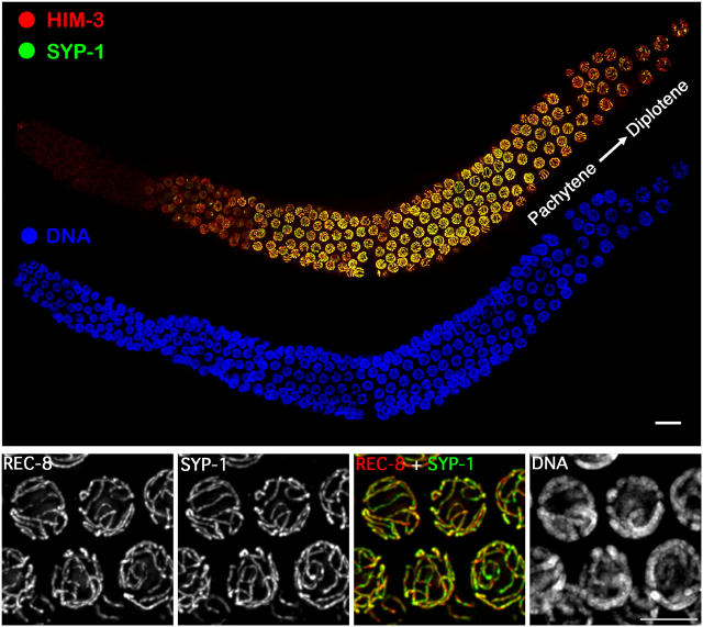
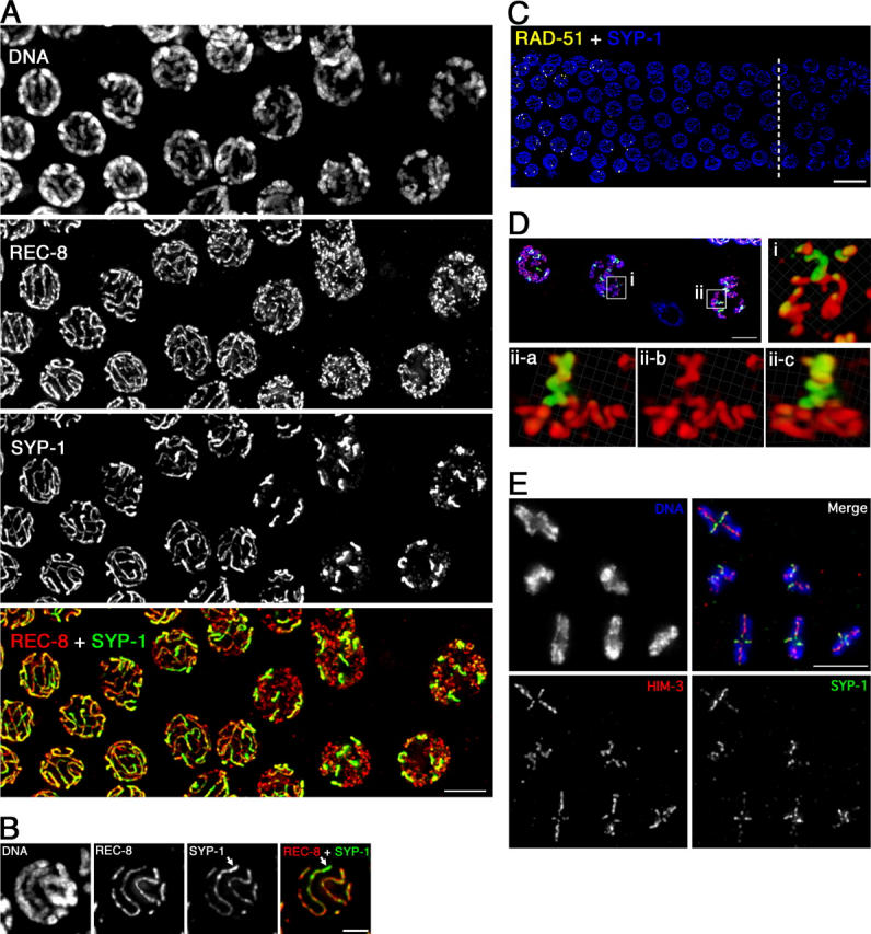
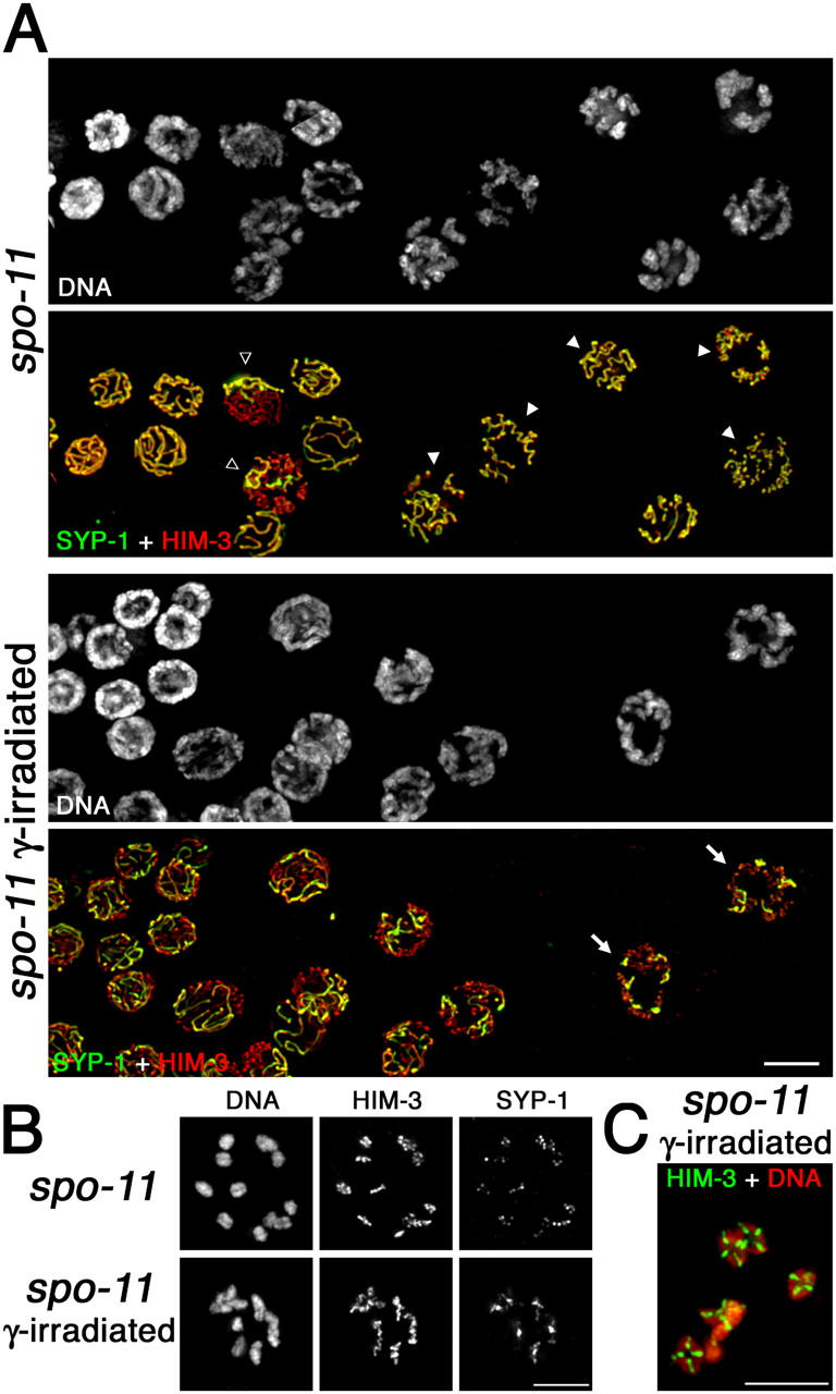
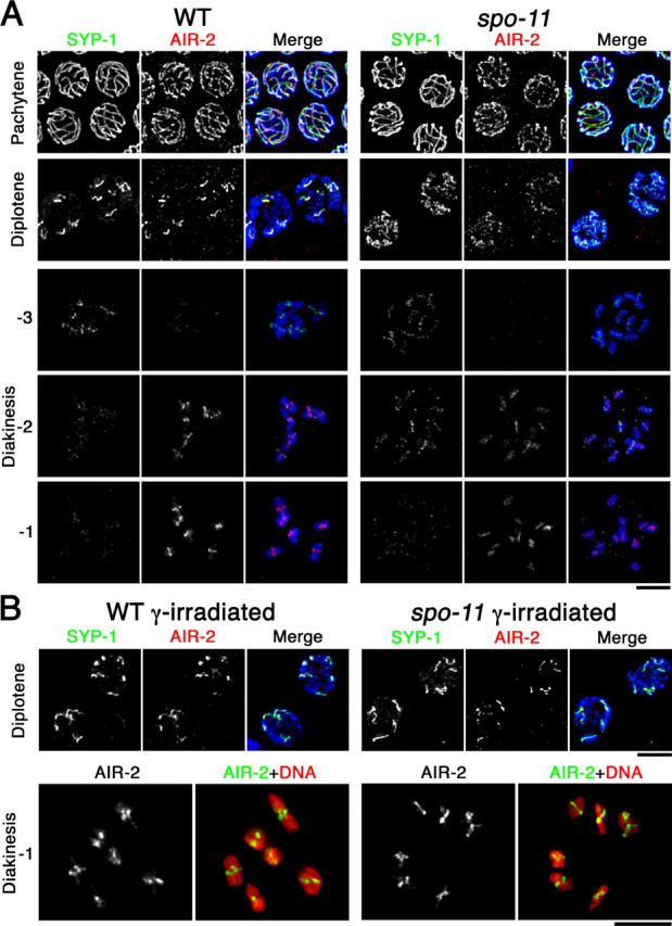
Homologous chromosome pairs (bivalents) undergo restructuring during meiotic prophase to convert a configuration that promotes crossover recombination into one that promotes bipolar spindle attachment and localized cohesion loss. We have imaged remodeling of meiotic chromosome structures after pachytene exit in Caenorhabditis elegans. Chromosome shortening during diplonema is accompanied by coiling of chromosome axes and highly asymmetric departure of synaptonemal complex (SC) central region proteins SYP-1 and SYP-2, which diminish over most of the length of each desynapsing bivalent while becoming concentrated on axis segments distal to the single emerging chiasma. This and other manifestations of asymmetry along chromosomes are lost in synapsis-proficient crossover-defective mutants, which often retain SYP-1,2 along the full lengths of coiled diplotene axes. Moreover, a gamma-irradiation treatment that restores crossovers in the spo-11 mutant also restores asymmetry of SYP-1 localization. We propose that crossovers or crossover precursors serve as symmetry-breaking events that promote differentiation of subregions of the bivalent by triggering asymmetric disassembly of the SC.


Figures

Figure 1.
Figure 1.
Immunolocalization of axis and SC components during wild-type meiosis. (Top) Whole mount germ line oriented with the distal premeiotic region at the left and the region of the pachytene–diplotene transition at the right. Bar, 10 μm. (Bottom) Pachytene nuclei exhibiting essentially full colocalization of SC central region protein SYP-1 and axis protein REC-8 between parallel tracks of DAPI-stained chromatin. Bar, 5 μm.
Figure 2.
Figure 2.
Chromosome reorganization after pachytene exit. See text for description. (A) Immunolocalization of REC-8 and SYP-1 at the pachytene–diplotene transition; late pachytene nuclei are at the left, diplotene at the right. Bar, 5 μm. (B) Projection halfway through a late pachytene nucleus showing unevenness in intensity of SYP-1 signal along bivalent (arrow). Bar, 2 μm. (C) Position of nuclei showing SYP-1 asymmetry (right side of a dashed line) relative to disappearance of RAD-51 foci. Bar, 10 μm. (D) Volume renderings of individual diplotene bivalents. Red, α-REC-8; green, α-SYP-1; blue, DAPI. i, X-shaped bivalent; ii-a–c, Y-shaped bivalent shown from two different angles. Bar, 5 μm. Videos 1 and 2 (available at http://www.jcb.org/cgi/content/full/jcb200410144/DC1) show rotating images of these bivalents. (E) Complete complement of bivalents from a single oocyte nucleus at early/mid-diakinesis. Bar, 5 μm.
Figure 3.
Figure 3.
Immunolocalization of SYP-2 after pachytene exit. α-SYP-2 staining and DAPI-stained chromatin in wild-type, spo-11, and msh-5 germ lines; portions shown extend from the region of pachytene exit (left) through late diplonema/early diakinesis (right). *, somatic nucleus. Bar, 10 μm.
Figure 4.
Figure 4.
Localization of SYP-1 and HIM-3 after pachytene exit in untreated and γ-irradiated spo-11 mutants. (A) Portions of germ lines extend from late pachynema (left) through mid/late diplonema (right). Closed arrowheads indicate diplotene nuclei in which SYP-1 is retained along the lengths of coiled axes; open arrowheads indicate nuclei in which SYP-1 has been lost from the majority of the chromosomes but is retained on some chromosomes; arrows indicate nuclei in which each desynapsing bivalent retains a single robust stretch of SYP-1, reflecting restoration of orderly asymmetric departure of SYP-1 by γ-irradiation. (B, top) Mid-diakinesis oocyte from an untreated spo-11 worm showing lack of chiasmata and SYP-1 distributed along the lengths of the axes. (bottom) Late diplotene oocyte from a γ-irradiated spo-11 worm showing SYP-1 concentrated on a region of each bivalent in the vicinity of and/or distal to the emerging chiasma. (C) Late diakinesis oocyte from a γ-irradiated spo-11 worm showing restoration of chiasmata. Bars, 5 μm.
Figure 5.
Figure 5.
Dynamic localization of SYP-1 and AIR-2. (A) Immunolocalization of SYP-1 and AIR-2 in wild-type and spo-11 nuclei at the indicated stages. For diakinesis nuclei, −3, −2, and −1 indicate position relative to the spermatheca, with the oocyte in the −1 position (closest to the spermatheca) being the most mature. In A and B, detection thresholds for α-AIR-2 signals were lower for the pachytene and diplotene panels than for the diakinesis panels to permit imaging of both the lower levels of chromosome-associated AIR-2 at the earlier stages and the subchromosomal localization of the higher levels of AIR-2 in late diakinesis. DAPI-stained chromatin is shown in blue in the merged images. (B) SYP-1 and AIR-2 localization after γ-irradiation. Bars, 5 μm.

References

    1. Albertson, D.G., A.M. Rose, and A.M. Villeneuve. 1997. Chromosome organization, mitosis, and meiosis. C. elegans II. D.L. Riddle, T. Blumenthal, B.J. Meyer, and J.R. Priess, editors. Cold Spring Harbor Laboratory Press, Plainview, NY. 47–78. - PubMed
    1. Blower, M.D., B.A. Sullivan, and G.H. Karpen. 2002. Conserved organization of centromeric chromatin in flies and humans. Dev. Cell. 2:319–330. - PMC - PubMed
    1. Chan, R.C., A.F. Severson, and B.J. Meyer. 2004. Condensin restructures chromosomes in preparation for meiotic divisions. J. Cell Biol. 167:613–625. - PMC - PubMed
    1. Colaiácovo, M.P., A.J. MacQueen, E. Martinez-Perez, K. McDonald, A. Adamo, A. La Volpe, and A.M. Villeneuve. 2003. Synaptonemal complex assembly in C. elegans is dispensable for loading strand-exchange proteins but critical for proper completion of recombination. Dev. Cell. 5:463–474. - PubMed
    1. Dernburg, A.F., K. McDonald, G. Moulder, R. Barstead, M. Dresser, and A.M. Villeneuve. 1998. Meiotic recombination in C. elegans initiates by a conserved mechanism and is dispensable for homologous chromosome synapsis. Cell. 94:387–398. - PubMed

Publication types

MeSH terms