. 2005 Feb 28;168(5):789-99.
doi: 10.1083/jcb.200409028.

Regulation of the interaction between PIPKI gamma and talin by proline-directed protein kinases

Affiliations

Regulation of the interaction between PIPKI gamma and talin by proline-directed protein kinases

Sang Yoon Lee et al. J Cell Biol. .

Abstract

The interaction of talin with phosphatidylinositol(4) phosphate 5 kinase type I gamma (PIPKI gamma) regulates PI(4,5)P2 synthesis at synapses and at focal adhesions. Here, we show that phosphorylation of serine 650 (S650) within the talin-binding sequence of human PIPKI gamma blocks this interaction. At synapses, S650 is phosphorylated by p35/Cdk5 and mitogen-activated protein kinase at rest, and dephosphorylated by calcineurin upon stimulation. S650 is also a substrate for cyclin B1/Cdk1 and its phosphorylation in mitosis correlates with focal adhesion disassembly. Phosphorylation by Src of the tyrosine adjacent to S650 (Y649 in human PIPKI gamma) was shown to enhance PIPKI gamma targeting to focal adhesions (Ling, K., R.L. Doughman, V.V. Iyer, A.J. Firestone, S.F. Bairstow, D.F. Mosher, M.D. Schaller, and R.A. Anderson. 2003. J. Cell Biol. 163:1339-1349). We find that Y649 phosphorylation does not stimulate directly PIPKI gamma binding to talin, but may do so indirectly by inhibiting S650 phosphorylation. Conversely, S650 phosphorylation inhibits Y649 phosphorylation by Src. The opposite effects of the phosphorylation of Y649 and S650 likely play a critical role in regulating synaptic function as well as the balance between cell adhesion and cell motility.


Figures
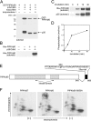
Figure 1.
Figure 1.
PIPKIγ90 is phosphorylated by p35/Cdk5 at S650 in vitro. Recombinant PIPKIγ90 fusion proteins were incubated with p35–Cdk5 complex in the presence of [32P]ATP for 30 min. Protein phosphorylation was analyzed by autoradiography after SDS-PAGE. (A) GST-PIPKIγ90 was phosphorylated by p35/Cdk5 in the absence and presence of the Cdk5 inhibitors, roscovitine or butyrolactone I (20 μM each). (B and C) Phosphorylation of His6-PIPKIγ90 by p35/Cdk5 and time course of the phosphorylation. (D) Stoichiometry of the phosphorylation of His6-PIPKIγ90 by p35/Cdk5. Purified proteins phosphorylated in vitro in the presence of [32P]ATP were separated by SDS-PAGE and stained with Coomassie brilliant blue. PIPKIγ90 bands were excised and incorporation of 32P was measured. (E) Diagram indicating the position of the phosphorylation site S650 in the COOH-terminal 28-aa tail of PIPKIγ90. (F) 2-D phosphopeptide mapping of His6-PIPKIγ87, His6-PIPKIγ90, and S650A mutant His6-PIPKIγ90 phosphorylated by p35/Cdk5 for 30 min in vitro. A circle outlines the spots present only in the protein that contains S650.
Figure 2.
Figure 2.
PIPKIγ90 is phosphorylated by p35/Cdk5 at S650 in vivo. (A) Generation and characterization of a polyclonal antibody specific for phospho-S650 (pS650). Purified WT and S650A mutant His6-PIPKIγ90 were incubated with or without p35/Cdk5 and with either nonradioactive ATP (for Western blotting) or [32P]ATP (for autoradiography) for 30 min. Samples were analyzed by autoradiography, and Western blotting with the anti-pS650 antibody was performed. Coomassie brilliant blue (CBB) staining shows loading of equal amounts of substrate proteins. The autoradiographic signal was quantified by a phosphorimager analysis and values were expressed as a percentage of the radioactivity incorporated into the WT sample. Bar graphs represent mean ± SD (n = 3). (B) CHO cells were transfected with WT or S650A mutant HA-PIPKIγ90 together with p35 and Cdk5. PIPKIγ90 immunoprecipitates were obtained from cell lysates 24 h after transfection. S650 phosphorylation and PIPKIγ90 expression were analyzed by Western blotting with anti-pS650 and anti-HA antibodies, respectively. (C) CHO cells were cotransfected with HA-PIPKIγ90, p35, and Cdk5 or mut-Cdk5. PIPKIγ90 immunoprecipitates from transfected and control cells were analyzed by Western blotting as described above. The faint band visible in nontransfected CHO cells may represent phosphorylated hamster PIPKIγ90. The pS650 immunoreactivity was quantified using an NIH image analysis software and shown as mean ± SD (n = 3).
Figure 3
Figure 3
Cdk5 phosphorylation of PIPKIγ90 inhibits its interaction with talin in vivo. (A) ITC analysis of the binding of 12-mer WT and pS650 peptides from the 28-aa tail of PIPKIγ90 to GST-talin head. Raw data as a function of time are shown in the top panels, and plots of the total heat released as a function of the molar ratio of each ligand are shown in the bottom panels. The continuous line in the bottom panels represents the nonlinear, least-squares best fits to the experimental data using a one-site model of binding. Note the roughly 1:1 stoichiometry indicated by the half-height point of the sigmoidal curve (bottom left; Turnbull and Daranas, 2003), and the complete absence of heat release in the case of the pS650 peptide. (B) CHO cells were cotransfected with HA-PIPKIγ90, p35, and Cdk5 or mut-Cdk5. Protein contents of the starting lysates were revealed by Western blotting. PIPKIγ90 and talin were immunoprecipitated from the cell lysates and presence of PIPKIγ90 and talin in each immunoprecipitate was detected by Western blotting.
Figure 4.
Figure 4.
Phosphomimetic mutation at S650 (S650D) disrupts the interaction of PIPKIγ90 with talin in vitro. (A) GST-F3 overlay assay. Nitrocellulose blots of WT and mutant His6-PIPKIγ90 were overlaid with or without GST-F3 fusion protein, and then overlaid with anti-GST antibody. CBB staining reveals equal load of the lanes. (B) Pull-down assay from rat brain extracts on bead-immobilized WT and mutant His6-PIPKIγ90. Bound talin was revealed by Western blotting, and equal amount of bait proteins was revealed by CBB staining. (C) The intensities of the GST-F3 and talin bands shown in A and B were quantified by an NIH image analysis software. Values from mutant proteins were normalized to that from WT and are represented as mean ± SD (n = 4). (D) Localization of transfected GFP-PIPKIγ90. NIH3T3 cells were transfected with GFP-PIPKIγ90 or its S650D mutant, and then were processed by immunofluorescence for vinculin immunoreactivity. Bar, 10 μm. (E) Pull-down assay from lysates of CHO cells transfected with WT and S650D mutant GFP-PIPKIγ90. Bead-immobilized GST-integrin β1 tail was used as a bait. Talin and PIPKIγ90 in the lysates or bead fractions were detected by Western blotting. (F) NIH3T3 cells transfected with mutant (S650N) GFP-PIPKIγ90 were immunostained with antivinculin antibody. Bar, 10 μm. (G) CHO cells were transfected with WT, S650D, or S650N GFP-PIPKIγ90. After 24 h of transfection, cell lysates were immunoprecipitated with anti-PIPKIγ90 antibody, and the presence of talin and PIPKIγ90 in the immunoprecipitates was analyzed by Western blotting.
Figure 5.
Figure 5.
Inhibitory effects of p35/Cdk5 overexpression on focal adhesion. NIH3T3 cells were cotransfected with both p35 and Cdk5 for 24 h, and the overexpressed proteins were detected by immunofluorescence microscopy with the antibodies indicated. Focal adhesions were visualized by antivinculin immunostaining, and actin was visualized by fluorescent phalloidin. Bar, 10 μm.
Figure 6.
Figure 6.
Phosphorylation of either Y649 or S650 inhibits the phosphorylation of the adjacent site. (A) Binding of PIPKIγ90 peptides to GST-talin head as determined by ITC. Note that both the dissociation constant (K d) and the enthalpy (ΔH) of the binding are similar for WT and pY649 peptides. The pY654 peptide has lower affinity, but the pS650 peptide did not bind. N/A, not available. (B) In vitro phosphorylation by Cdk5 and c-Src of WT, pY649 and pS650 12-mer PIPKIγ90 peptides. Each peptide was incubated in the presence of [32P]ATP and of either p35/Cdk5 or c-Src for 20 min at 30°C. Peptides were then recovered on phosphocellulose paper, and the associated radioactivity was measured by Cerenkov counting. Incorporation of radioactivity into pY649 and pS650 peptides was normalized to that of the WT peptide and presented as mean ± SEM (n = 4)
Figure 7.
Figure 7.
Regulation of S650 phosphorylation in synaptosomes. In all panels shown in the figure, with the exception of E and F, levels of total PIPKIγ90 and of its pS650 epitope were analyzed by Western blots of anti-PIPKIγ90 immunoprecipitates obtained from synaptosomal lysates. For other proteins, Western blots were performed directly on synaptosomal lysates. (A) Freshly prepared rat brain synaptosomes, or synaptosomes exposed to a 1-h labeling step with 32Pi, were incubated for 1 min with either control buffer or stimulation buffer (high K+) in the absence or presence of Ca2+ (Bauerfeind et al., 1997). Anti-PIPKIγ90 immunoprecipitates prepared from these samples were analyzed by autoradiography (32P-labeled samples) or by Western blotting for pS650 and total PIPKIγ90. Western blotting for amphiphysin 2 revealed the previously described stimulation-dependent mobility shift of the upper band (because of its dephosphorylation), thus confirming the occurrence of Ca2+-dependent stimulation. (B) Synaptosomes were stimulated with high K+ for 1 min in the absence or presence of 2 μM cyclosporin A, and then were analyzed with antibodies directed against pS650 or phospho-sites 4 and 5 (MAPK sites) of synapsin I. (C) Synaptosomes were exposed for 1 min to either control buffer (resting) or high K+ buffer (depolarization). Aliquots of stimulated synaptosomes were then returned for 15 min to control buffer (repolarization) with and without the Cdk5 inhibitor butyrolactone I (10 μM). (D) Synaptosomes were incubated for 1 min with control buffer or high K+ buffer in the absence or presence of 1 μM okadaic acid, and then were analyzed for levels of pS650, PIPKIγ90, phospho-MAPK1/2, and total MAPK1/2. (E) Synaptosomes were stimulated for 1 min in the absence or presence of 1 μM okadaic acid and the MAPK inhibitor PD98059. (F) In vitro phosphorylation of WT and S650A mutant His6-PIPKIγ90 with purified MAPK1. S650 phosphorylation by MAPK1 was detected by the anti-pS650 antibody.
Figure 8.
Figure 8.
S650 of PIPKIγ90 undergoes mitotic phosphorylation by cyclin B1/Cdk1. (A) CHO cells transfected with WT or S650A mutant HA-PIPKIγ90 for 24 h were arrested in the mitotic state (M) by nocodazole treatment. G1 interphase cells (G) were further prepared from mitotically synchronized cells after removal of nocodazole. Cell lysates of both M and G cells were analyzed by Western blotting with anti-pS650 and anti-PIPKIγ90 antibodies. (B) U87MG cells were processed as described in A to generate mitotic and interphase cells. PIPKIγ90 was immunoprecipitated from cell lysates for analysis of pS650, talin and total PIPKIγ90. Cell lysates were also analyzed by Western blotting for levels of histone H3 phosphorylation using a phosphospecific antibody. Bar graphs represent normalized talin immunoreactivity (mean ± SD; n = 4) after quantification as shown in Fig. 4 C. (C) WT and S650A mutant His6-PIPKIγ90 were incubated in vitro with or without the cyclin B1–Cdk1 complex in the presence of [32P]ATP. His6-PIPKIγ90 phosphorylation and cyclin B1 autophosphorylation were examined by autoradiography after SDS-PAGE. In parallel, samples phosphorylated under the same conditions with nonradioactive ATP were processed for Western blotting with anti-pS650 antibody. CBB staining demonstrates equal amounts of proteins.

References

    1. Aikawa, Y., and T.F. Martin. 2003. ARF6 regulates a plasma membrane pool of phosphatidylinositol(4,5)bisphosphate required for regulated exocytosis. J. Cell Biol. 162:647–659. - PMC - PubMed
    1. Ajiro, K., K. Yoda, K. Utsumi, and Y. Nishikawa. 1996. Alteration of cell cycle-dependent histone phosphorylations by okadaic acid. Induction of mitosis-specific H3 phosphorylation and chromatin condensation in mammalian interphase cells. J. Biol. Chem. 271:13197–13201. - PubMed
    1. Alessi, D.R., N. Gomez, G. Moorhead, T. Lewis, S.M. Keyse, and P. Cohen. 1995. Inactivation of p42 MAP kinase by protein phosphatase 2A and a protein tyrosine phosphatase, but not CL100, in various cell lines. Curr. Biol. 5:283–295. - PubMed
    1. Barsukov, I.L., A. Prescot, N. Bate, B. Patel, D.N. Floyd, N. Bhanji, C.R. Bagshaw, K. Letinic, G. Di Paolo, P. De Camilli, et al. 2003. Phosphatidylinositol phosphate kinase type 1γ and β1-integrin cytoplasmic domain bind to the same region in the talin FERM domain. J. Biol. Chem. 278:31202–31209. - PubMed
    1. Bauerfeind, R., K. Takei, and P. De Camilli. 1997. Amphiphysin I is associated with coated endocytic intermediates and undergoes stimulation-dependent dephosphorylation in nerve terminals. J. Biol. Chem. 272:30984–30992. - PubMed

Publication types

MeSH terms