. 2005 Mar 8;102(10):3834-9.
doi: 10.1073/pnas.0500748102. Epub 2005 Feb 28.

Function and regulation of Tumbleweed (RacGAP50C) in neuroblast proliferation and neuronal morphogenesis

Affiliations

Function and regulation of Tumbleweed (RacGAP50C) in neuroblast proliferation and neuronal morphogenesis

Ann Y N Goldstein et al. Proc Natl Acad Sci U S A. .

Abstract

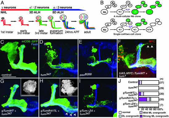
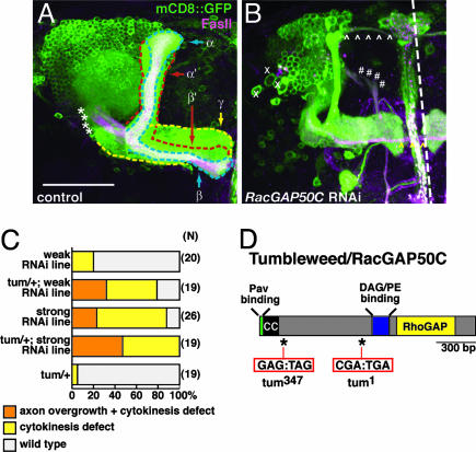
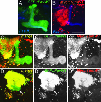
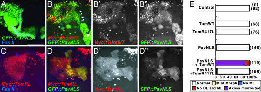
Drosophila RacGAP50C and its homologues act as part of a complex with a kinesin-like protein (Pavarotti/Zen-4) that is essential for the formation of the central spindle and completion of cytokinesis [Mishima, M., Kaitna, S. & Glotzer, M. (2002) Dev. Cell 2, 41-54; Somers, W. G. & Saint, R. (2003) Dev. Cell 4, 29-39; Jantsch-Plunger et al. (2000) J. Cell Biol. 149, 1391-1404]. We report here that RacGAP50C corresponds to the tumbleweed (tum) gene previously identified based on its defects in dendrite development of sensory neurons [Gao, F. B., Brenman, J. E., Jan, L. Y. & Jan, Y. N. (1999) Genes Dev. 13, 2549-2561]. Using mushroom body neurogenesis and morphogenesis as a model, we show that Tumbleweed (Tum), Pavarotti, and their association are required for neuroblast proliferation. Tum with a mutation predicted to disrupt the GTPase-activating protein (GAP) activity still largely retains its activity in regulating cell division but is impaired in its activity to limit axon growth. We also provide evidence that Tum and Pavarotti regulate the subcellular localization of each other in postmitotic neurons and that cytoplasmic accumulation of both proteins disrupts axon development in a GAP-dependent manner. Taken together with previous studies of RacGAP50C in regulating cytokinesis, we propose that Tum serves as a scaffolding protein in regulating cell division but acts as a GAP to limit axon growth in postmitotic neurons.


Figures

Fig. 2.
Fig. 2.
MARCM analysis of tum and pav in MB Nbs. (A) Timeline of MB neuron generation (adapted from ref. 19). NHL, newly hatched larvae; ALH, after larval hatching; APF, after puparium formation. (B) Generation of marked and homozygous mutant Nb vs. single-neuron (N) clone depends on FLP recombinase (FLP) activation in Nb or in the ganglion mother cell (G). (C) Control Nb clone generated in newly hatched larva. (D and E) tum347 and pavB200 Nb clone generated at the same stage. Xs mark presence of enlarged cell bodies, and severe decrease of cells generated from the Nb leads to the absence of all but the axons of the earliest born γ neurons. FasII highlights the axon projections of heterozygous MB neurons generated from the three other MB Nbs. (F) tum1 Nb clone expressing UAS-MYC::Tum cDNA partially rescues the cytokinetic phenotype, allowing for the generation of most but not all of the later-born neurons. Arrowheads mark axons that overextend beyond the normal dorsal lobe. (G) A transgene encompassing genomic WT tum (gTumWT) rescues tum347 Nb clone phenotypes. (Inset) Cell bodies from the same Nb and the absence of enlarged cell bodies. (H) A transgene for genomic tum with the R417L mutation also rescues proliferation defects in the tum347 Nb clone. (Inset) Cell bodies from same Nb. Arrowheads mark axons extending across the midline beyond normal ML. (I) gTum with a deletion in the Pav binding domain (ΔZ4) does not rescue Nb proliferation defect of tum347 clone. (J) Quantification of Nb clones that exhibit overextension beyond their normal lobe boundaries. (Scale bar: 50 μm.)
Fig. 1.
Fig. 1.
RNAi phenotypes of RacGAP50C/Tum. (A) WT MB complex labeled by GAL4-OK107 driven mCD8::GFP expression. Midline of the brain is at the right side of the image and dorsal is at the top in this and all subsequent images. The MB axons are organized in lobes outlined as: γ lobe (yellow), α′/β′ lobe (red), and α/β lobe (blue). These lobes are also distinguished by their expression of FasII (magenta). The α and α′ lobes are referred to collectively as the dorsal lobe (DL). The medial lobe (ML) includes the γ, β, and β′ lobes. Asterisks demarcate the peduncle. (Scale bar: 50 μm.) (B) Representative image illustrating phenotypes seen with RacGAP50C RNAi in the MB. Xs mark enlarged cell bodies; *caret;s follow the projection of axons overextending from the DL (white ⁁) and the ML (yellow ⁁). #s mark axons that bypass the peduncle and emerge in a medial trajectory from the dendritic region. (C) Quantitative analysis of RacGAP50C RNAi phenotypes. Heterozygous background of tum enhances MB phenotypes seen with weak and strong transgenic RacGAP50C RNAi lines. (D) Sequence analysis of two independently generated tum chromosomes revealed that they both contain nonsense mutations (tum1:R281STOP and tum347:E114STOP) in the coding region of RacGAP50C. CC, coiled-coil domain.
Fig. 3.
Fig. 3.
Tum and Pav influence each other's localization, and their association leads to disruption of axon routing. (A) UAS-GFP::PavWT is detected in the axons as well as the nuclei of MB neurons when driven by GAL4-OK107. (B) UAS-MYC::TumWT is restricted to the nuclei of MB neurons. (C and D) Two examples of coexpression of UAS-GFP::PavWT and UAS-MYC::Tum leading to their colocalizations. C and D are merged signals for GFP (C′ and D′) and MYC (C′′ and D′′). Example in D shows that coexpression leads to axon misrouting that forms a ball-like shape close to the cell body region of the MB complex (arrow). (Scale bar: 50 μm.)
Fig. 4.
Fig. 4.
Tum/Pav coexpression phenotype is enhanced by increased cytoplasmic concentration and abolished by a mutation in the RhoGAP domain. (A) GFP::PavNLS is excluded from the nucleus and detected in the axons and dendrites of MB neurons. (BB′′) Coexpression of UAS-GFP::PavNLS and UAS-MYC::Tum leads to their overlapping subcellular localization. Arrowheads follow misrouted axons that bypass the peduncle (*) and misguide to a region ventral to the medial lobe (#) before innervating part of the medial lobe. B is a merge of MYC (B′) and GFP (B″) signals. (C) MYC::TumR417L is restricted to the nuclei of MB neurons when expressed alone. (DD′′) Coexpression of UAS-GFP::PavNLS and UAS-MYC::TumR417L leads to a similar overlap of their subcellular localization but does not lead to a gross change in the axonal targeting of MB neurons. D is a merge of MYC (D′) and GFP (D″) signals. (Scale bar: 50 μm.) (E) Quantification of axon misrouting with the expression of Tum and Pav transgenes. DL, dorsal lobe; ML, medial lobe. See Materials and Methods for details of analysis.

References

    1. Scheffzek, K., Ahmadian, M. R., Kabsch, W., Wiesmuller, L., Lautwein, A., Schmitz, F. & Wittinghofer, A. (1997) Science 277, 333-338. - PubMed
    1. Lander, E. S. (2001) Nature 409, 860-921. - PubMed
    1. Venter, J. C. (2001) Science 291, 1304-1351. - PubMed
    1. Endris, V., Wogatzky, B., Leimer, U., Bartsch, D., Zatyka, M., Latif, F., Maher, E. R., Tariverdian, G., Kirsch, S., Karch, D. & Rappold, G. A. (2002) Proc. Natl. Acad. Sci. USA 99, 11754-11759. - PMC - PubMed
    1. Billuart, P., Bienvenu, T., Ronce, N., des Portes, V., Vinet, M. C., Zemni, R., Roest Crollius, H., Carrie, A., Fauchereau, F., Cherry, M., et al. (1998) Nature 392, 923-926. - PubMed

Publication types

Substances

LinkOut - more resources