. 2005 Mar 8;102(10):3756-9.
doi: 10.1073/pnas.0409910102. Epub 2005 Feb 28.

Culturing at atmospheric oxygen levels impacts lymphocyte function

Affiliations

Culturing at atmospheric oxygen levels impacts lymphocyte function

Kondala R Atkuri et al. Proc Natl Acad Sci U S A. .

Abstract

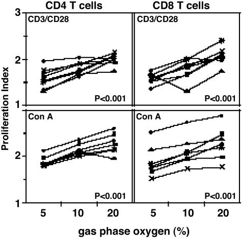
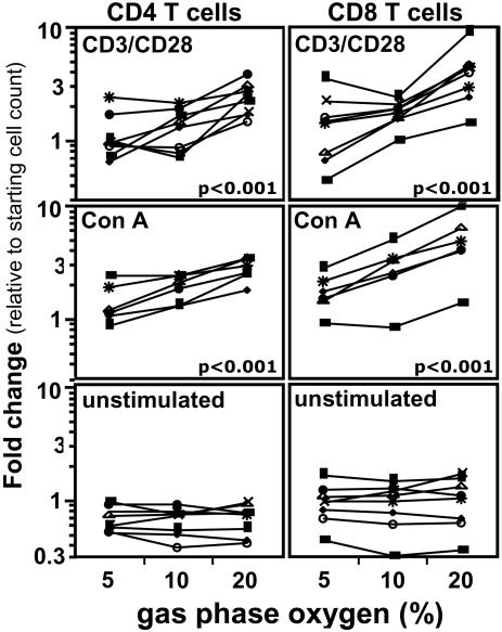
To determine whether culturing peripheral blood mononuclear cells at atmospheric oxygen levels skews responses in comparison with culturing lymphocytes at physiologic oxygen levels, we cultured peripheral blood mononuclear cells at 5%, 10%, and atmospheric (20%) gas-phase oxygen for 5 days. We found that incubator oxygen levels influenced lymphocyte proliferation stimulated by two commonly used stimuli: Con A and antibodies that crosslink surface CD3 and CD28 to mimic antigen presentation. In both cases, proliferation increased as gas-phase oxygen levels increased. In contrast, oxygen levels did not influence proliferation stimulated by phytohemagglutinin, another commonly used mitogen. Similarly, oxygen levels did not impact cell viability in unstimulated cultures. Thus, we conclude that the influence of oxygen levels on proliferation depends on the stimulus, and, most importantly from the standpoint of immune responses, culturing cells at atmospheric rather than physiologic oxygen levels results in significantly increased proliferation responses to the CD3/CD28 crosslinking, a proliferation stimulus commonly used to mimic T cell antigen receptor signaling.


Figures

Fig. 1.
Fig. 1.
Proliferation indices of human lymphocytes stimulated with CD3/CD28 crosslinking and Con A are higher at 20% oxygen. Human PBMC stained with CFDA-SE were cultured at 5%, 10%, and 20% oxygen for 5 days (see Materials and Methods). Shown are lymphocytes stimulated by CD3/CD28 crosslinking (Upper) and with Con A (20 μg/ml, Lower). Statistics were calculated by using jmp software by least-square-fit model with sample and oxygen as independent variables (see Materials and Methods). Each set of connected points represents one subject (n = 8).
Fig. 2.
Fig. 2.
Increase in total number of CD4 and CD8 T cells stimulated with CD3/CD28 crosslinking and Con A is higher at 20% oxygen. Human PBMC stained with CFDA-SE were stimulated at 5%, 10%, and 20% oxygen for 5 days (see Materials and Methods). Shown are cells stimulated with CD3/CD28 crosslinking (Top), cells stimulated with Con A (20 μg/ml, Middle), and unstimulated cells (Bottom). Cell counts were performed by using BD Trucount beads (see Materials and Methods). Fold change is calculated for each subject for CD4 and CD8 T cell subsets as the ratio of the number of live cells in the subset at day 5 to the number of live cells in the subset at the beginning of the culture. Statistics were calculated using jmp software by least-square-fit model with sample and oxygen as independent variables (see Materials and Methods). Each set of connected points represents one subject (n = 6). The numbers of live CD4 and CD8 T cells present in unstimulated cultures decreased 0.3-fold on average by the end of the culture period (e.g., CD4 T cells for a typical subject decreased from 3.6 × 10⁵ cells at the start to 2.6 × 10⁵ cells at the end of the culture period). This decrease was independent of the incubator oxygen tension (P > 0.05).

References

    1. Steurer, J., Hoffmann, U., Dur, P., Russi, E. & Vetter, W. (1997) Respiration 64, 200-205. - PubMed
    1. Chow, D. C., Wenning, L. A., Miller, W. M. & Papoutsakis, E. T. (2001) Biophys. J. 81, 675-684. - PMC - PubMed
    1. Krieger, J. A., Landsiedel, J. C. & Lawrence, D. A. (1996) Int. J. Immunopharmacol. 18, 545-552. - PubMed
    1. Caldwell, C. C., Kojima, H., Lukashev, D., Armstrong, J., Farber, M., Apasov, S. G. & Sitkovsky, M. V. (2001) J. Immunol. 167, 6140-6149. - PubMed
    1. Derevianko, A., D'Amico, R. & Simms, H. (1996) Clin. Exp. Immunol. 106, 560-567. - PMC - PubMed

Publication types

LinkOut - more resources