. 2005 Mar 8;102(10):3605-10.
doi: 10.1073/pnas.0409889102. Epub 2005 Feb 28.

Regulation of heterogenous nuclear ribonucleoprotein A1 transport by phosphorylation in cells stressed by osmotic shock

Affiliations

Regulation of heterogenous nuclear ribonucleoprotein A1 transport by phosphorylation in cells stressed by osmotic shock

Eric Allemand et al. Proc Natl Acad Sci U S A. .

Abstract

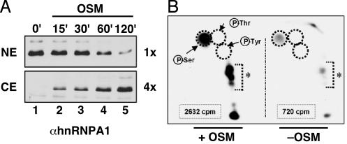
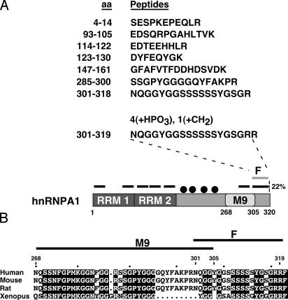
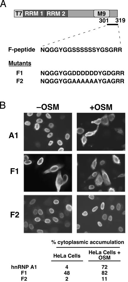
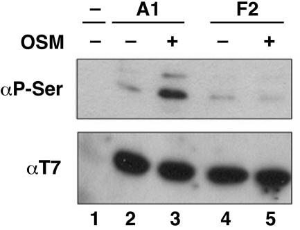
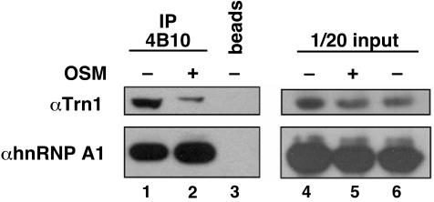
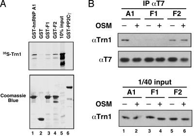
Heterogeneous nuclear ribonucleoprotein (hnRNP) A1 is an alternative splicing factor that is mainly nuclear, although it shuttles rapidly between nuclear and cytoplasmic compartments. Cells stressed by osmotic shock (OSM) activate the mitogen-activated protein kinase kinase(3/6)-p38 signaling pathway, which in turn results in accumulation of hnRNP A1 in the cytoplasm. This effect modulates alternative splicing regulation in vivo and correlates with increased hnRNP A1 phosphorylation. We have characterized the molecular mechanism involved in the cytoplasmic accumulation of hnRNP A1 in NIH 3T3 cells subjected to OSM. This treatment results in serine-specific phosphorylation within a C-terminal peptide, dubbed the "F-peptide," which is adjacent to the M9 motif that mediates bidirectional transport of hnRNP A1. Analysis of mutants in which the F-peptide serines were replaced by aspartic acids or alanines showed that F-peptide phosphorylation is required for the subcellular redistribution of hnRNP A1 in cells subjected to OSM. Furthermore, F-peptide phosphorylation modulates the interaction of hnRNP A1 with transportin Trn1. Our findings suggest that the phosphorylation of F-peptide by cell-signaling pathways regulates the rate of hnRNP A1 nuclear import.


Figures

Fig. 1.
Fig. 1.
Subcellular distribution and specific phosphorylation of hnRNP A1 in cells subjected to OSM. (A) Nuclear (NE) and cytoplasmic (CE) extracts were prepared from NIH 3T3 cells treated with 600 mM sorbitol for the indicated times. The distribution of endogenous hnRNP A1 was detected by Western blotting with mAb A1/55 by using a 4-fold excess of CE over NE total protein. (B) Phosphoamino acid analysis of metabolically labeled hnRNP A1 by 2D electrophoresis after immunoprecipitation from cells treated with OSM or untreated. Equal amounts of hnRNP A1 (estimated by Ponceau red staining) were excised, quantitated by Cerenkov counting (indicated for each condition), and processed for phosphoamino acid analysis. The positions of phosphoserine (P-Ser), phosphotyrosine (P-Tyr), or phosphothreonine (P-Thr) standards are shown as dotted circles. *, Partially digested peptides.
Fig. 2.
Fig. 2.
hnRNP A1 peptides identified by MALDI-TOF. (A) The identified peptides are listed with their coordinates in the 320-aa hnRNP A1 sequence. The positions of the seven peptides also are shown as black bars over a diagram of the hnRNP A1 protein, and the modified F-peptide is shown as a gray bar. The four black dots represent the methylated arginines R193, R205, R217, and R224. (B) Comparative alignment of the M9 sequence (268–305) and the F-peptide (301–319). The C termini of several vertebrate hnRNP A1 orthologs were aligned by using clustalw (www.ebi.ac.uk/clustalw). Residues identical in all four species are highlighted.
Fig. 3.
Fig. 3.
Localization of transfected wild-type or mutant hnRNP A1. (A) The domain structure of T7-tagged hnRNP A1 and F1/F2 mutants is shown schematically. The mutations of serines (S308–S313 and S316) to aspartic acids (F1) or alanines (F2) in the F-peptide are shown. (B) Immunofluorescence micrographs showing the localization of T7-hnRNP A1, T7-F1, and T7-F2 in HeLa cells, with or without OSM treatment for 2.5 h. The tabulated data indicate the percentage of transfected cells in which the tagged proteins accumulated in the cytoplasm. Approximately 500 cells were counted for each condition.
Fig. 4.
Fig. 4.
Mutation of the F-peptide's seven serines to alanines abolishes the increase of hnRNP A1 phosphorylation in cells subjected to OSM. HeLa cells were transfected with T7-hnRNP A1 or T7-F2 plasmids and treated with OSM for 3 h. The transiently expressed proteins were immunoprecipitated from total protein lysates by using a T7-tag mAb and separated by SDS/PAGE. The levels of serine phosphorylation and immunoprecipitated proteins were determined by Western blotting with anti-phosphoserine and anti-T7 antibodies, respectively.
Fig. 5.
Fig. 5.
Decreased association of hnRNP A1 with Trn1 in OSM-stressed cells. hnRNP A1 was immunoprecipitated from cytoplasmic extracts with mAb 4B10. The immunoprecipitated proteins were separated by SDS/PAGE, and Trn1 and hnRNP A1 were detected by Western blotting with D45 and 4B10 antibodies, respectively. In the panels on the right, 1/20th of the amount of each protein extract used for immunoprecipitation was analyzed as a control for protein expression level.
Fig. 6.
Fig. 6.
Hyperphosphorylation of F-peptide reduced the interaction of hnRNP A1 with Trn1. (A) The indicated GST recombinant proteins were bound to gluthatione-Sepharose and assayed for binding to in vitro-translated [35S]-Trn1; The proteins were analyzed by SDS/PAGE (Coomassie blue; Lower), and [35S]-Trn1 was detected by using autoradiography ([35S]-Trn1; Upper). (B) T7 plasmids expressing hnRNP A1 or F1/F2 mutants were transfected into HeLa cells, and the transiently expressed proteins were immunoprecipitated from total protein lysates with a T7-tag mAb. The T7-tagged proteins and the coimmunoprecipitated endogenous Trn1 were detected by Western blotting with T7 and D45 antibodies, respectively. A fraction of the total protein lysate (1/40th of input) also was analyzed to determine the level of endogenous Trn1 expressed under each condition.

Similar articles

Cited by

References

    1. Dreyfuss, G., Kim, V. N. & Kataoka, N. (2002) Nat. Rev. Mol. Cell Biol. 3, 195-205. - PubMed
    1. Krecic, A. M. & Swanson, M. S. (1999) Curr. Opin. Cell Biol. 11, 363-371. - PubMed
    1. Piñol-Roma, S., Choi, Y. D., Matunis, M. J. & Dreyfuss, G. (1988) Genes Dev. 2, 215-227. - PubMed
    1. Dreyfuss, G., Matunis, M. J., Piñol-Roma, S. & Burd, C. G. (1993) Annu. Rev. Biochem. 62, 289-321. - PubMed
    1. Michael, W. M., Siomi, H., Choi, M., Piñol-Roma, S., Nakielny, S., Liu, Q. & Dreyfuss, G. (1995) Cold Spring Harb. Symp. Quant. Biol. 60, 663-668. - PubMed

Publication types

MeSH terms

Substances

LinkOut - more resources