. 2005 Mar 8;102(10):3782-7.
doi: 10.1073/pnas.0408310102. Epub 2005 Feb 28.

Regulation of platelet granule exocytosis by S-nitrosylation

Affiliations

Regulation of platelet granule exocytosis by S-nitrosylation

Craig N Morrell et al. Proc Natl Acad Sci U S A. .

Abstract

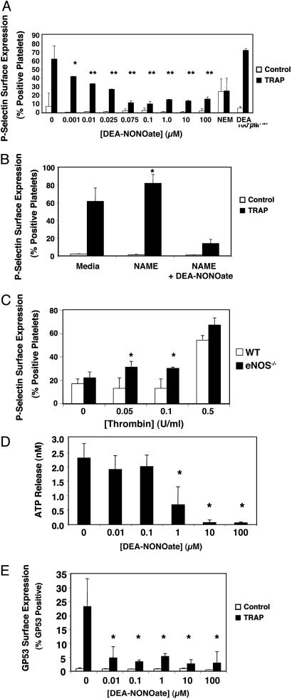
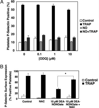
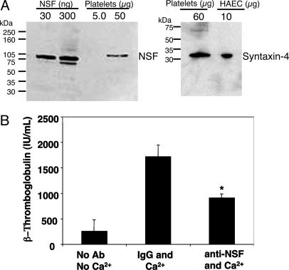
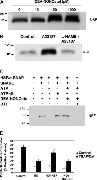
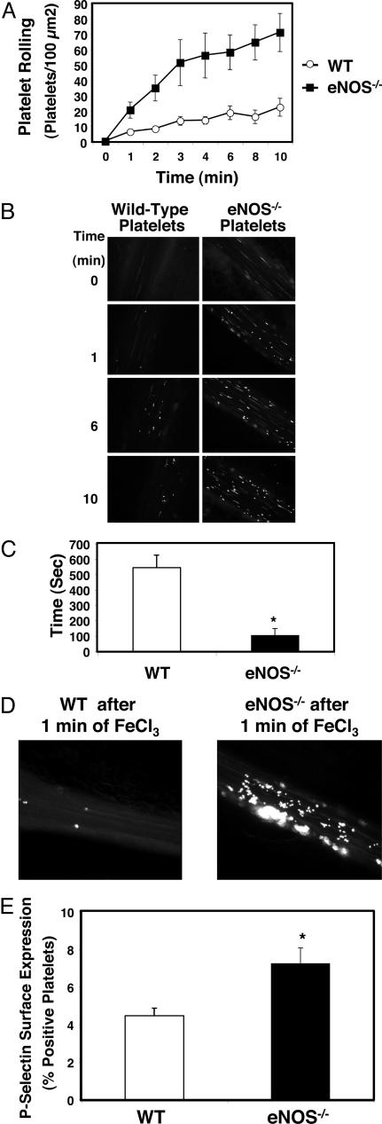
Nitric oxide (NO) regulates platelet activation by cGMP-dependent mechanisms and by mechanisms that are not completely defined. Platelet activation includes exocytosis of platelet granules, releasing mediators that regulate interactions between platelets, leukocytes, and endothelial cells. Exocytosis is mediated in part by N-ethylmaleimide-sensitive factor (NSF), an ATPase that disassembles complexes of soluble NSF attachment protein receptors. We now demonstrate that NO inhibits exocytosis of dense granules, lysosomal granules, and alpha-granules from human platelets by S-nitrosylation of NSF. Platelets lacking endothelial NO synthase show increased rolling on venules, increased thrombosis in arterioles, and increased exocytosis in vivo. Regulation of exocytosis is thus a mechanism by which NO regulates thrombosis.


Figures

Fig. 1.
Fig. 1.
NO inhibition of platelet granule exocytosis. (A) Exogenous NO inhibits α-granule exocytosis: P-selectin. Human platelets were incubated with DEA-NONOate for 15 min and treated with 15 μM TRAP, and the amount of P-selectin on the surface was measured by FACS. (n = 6 ± SD, *, P = 0.05; **, P < 0.01 vs. TRAP without NO donor.) (B) Endogenous NO inhibits α-granule exocytosis. Human platelets were pretreated with 1 mM l-NAME for 15 min to inhibit endogenous NOS and treated with 15 μM TRAP, and the amount of P-selectin on the surface was measured by FACS. (n = 3–6 ± SD, *, P < 0.01 vs. 0 mM l-NAME.) (C) Endogenous NO inhibits α-granule exocytosis. Platelets from wild-type and eNOS-/- mice were treated with control or thrombin, and the amount of P-selectin on the surface was measured by FACS. (n = 3 ± SD, *, P < 0.01 vs. WT.) (D) NO inhibits dense-granule exocytosis. Human platelets were pretreated with DEA-NONOate for 15 min and treated with 15 μM TRAP, and the amount of released ATP was measured by chemiluminescence. (n = 3 ± SD, *, P < 0.01 vs. 0 μM.) (E) NO inhibits lysosomal granule release. Human platelets were pretreated with increasing amounts of DEA-NONOate for 15 min and treated with 15 μM TRAP, and the amount of gp53 (CD63) on the surface was measured by FACS. (n = 3 ± SD, *, P = < 0.01 vs. 0 μM.)
Fig. 2.
Fig. 2.
NO inhibition of platelet α-granule exocytosis is independent of cGMP. (A) Inhibition of guanylate cyclase does not affect NO inhibition of exocytosis. Human platelets were pretreated with ODQ for 10 min, then treated with 10 μM DEA-NONOate for 15 min and finally treated with control or 15 μM TRAP, and the amount of P-selectin on the surface was measured by FACS. (n = 3 ± SD.) (B) The denitrosylating agent NAC reverses NO inhibition of platelet granule exocytosis. Human platelets were pretreated with 10 μM DEA-NONOate for 10 min, then treated with 1 mM N-acetyl-cysteine for 10 min and finally treated with control or 15 μM TRAP, and the amount of P-selectin on the surface was measured by FACS. (n = 3 ± SD.)
Fig. 3.
Fig. 3.
NSF regulates platelet granule exocytosis. (A) Human platelets contain NSF and syntaxin-4. Lysates of human platelets, recombinant NSF, or human aortic endothelial cells (HAEC) were immunoblotted with antibody to NSF (Left) or syntaxin-4 (Right). (B) NSF regulates α-granule release: antibody inhibition. Human platelets were permeabilized, incubated with antibody, and activated with Ca2+. β-Thromboglobulin release was measured by ELISA. (n = 4–6 ± SD, *, P < 0.05 vs. IgG and Ca2+.)
Fig. 4.
Fig. 4.
NO inhibits NSF regulation of platelet granule exocytosis. (A) NSF is a target of exogenous NO: dose–response. Murine platelets were harvested, and equal numbers were incubated with control or DEA-NONOate, lysed, immunoprecipitated with antibody to nitrosocysteine, and immunoblotted with antibody to NSF. NSF is nitrosylated in a dose-dependent manner. (Each step was repeated three times with similar results.) (B) NSF is a target of endogenous NO. Murine platelets were harvested, preincubated with control or l-NAME 5 mM, treated with control or 1 μM Ca2+ ionophore, lysed, immunoprecipitated with antibody to nitrosocysteine, and immunoblotted with antibody to NSF. (C) NO inhibits NSF separation from syntaxin-4 in vitro. Recombinant His6-NSF was pretreated or not pretreated with DEA-NONOate and incubated with a-SNAP and GST-SNARE fusion polypeptides expressed in platelets. ATP or ATP-gS was added, and the mixture was precipitated with glutathione-Sepharose. Precipitated proteins were immunoblotted with antibody to the NSF tag. (D) NSF is a target of NO. Platelets were permeabilized, exposed to 1 mM DEA-NONOate, and then incubated with recombinant NSF or nitrosylated NSF. Platelets were then stimulated or not stimulated with TRAP and Ca2+, and exocytosis was measured by FACS. (n = 3 ± SD, *, P < 0.05 for NO plus NSF vs. NO plus NSF-NO.)
Fig. 5.
Fig. 5.
NO inhibits platelet adherence and aggregation in vivo. (A) Platelet rolling. Wild-type mice were anesthetized and injected with calcein-AM-labeled platelets from wild-type (WT) or eNOS-/- mice. The mesentery was externalized, and venules 120–150 μm in diameter were treated with 250 mM FeCl3. Platelet rolling on venules was imaged with a digital fluorescent camera (n = 7–8 ± SEM). (B) Digital fluorescence images of platelet rolling. Wild-type mice were anesthetized and injected with calcein-AM-labeled platelets from wild-type or eNOS-/- mice. The mesentery was externalized, and venules 120–150 μm in diameter were treated with 250 mM FeCl3. Platelet rolling on venules was imaged with a digital fluorescent camera 0, 1, 6, and 10 min after FeCl3 treatment. (Representative images from n = 7–8.) (C) Thrombosis. Mice were injected with labeled platelets as above. The mesentery was externalized, and arterioles 50–80 μm in diameter were treated with 500 mM FeCl3. The time to formation of the first thrombus >10 μm in size was recorded (n = 5 ± SD). (D) Digital fluorescent images of thrombosis. Wild-type and eNOS-/- mice were injected with labeled wild-type platelets, arterioles were treated with FeCl3 as above, and a digital fluorescent camera was used to visualize formation of the first thrombus >10 μm in size. (E) Exocytosis in shed platelets. The distal 3 mm of wild-type and eNOS-/- mice tails were amputated, and shed blood was collected. Platelets were analyzed for surface P-selectin expression by FACS. Shed platelets from eNOS-/- have increased exocytosis as measured by P-selectin expression compared with WT mice. (n = 3 ± SD, *, P < 0.002 for WT vs. eNOS-/-.)

Similar articles

Cited by

References

    1. Moncada, S., Palmer, R. M. & Higgs, E. A. (1991) Pharmacol. Rev. 43, 109-142. - PubMed
    1. Loscalzo, J. (2001) Circ. Res. 88, 756-762. - PubMed
    1. Michel, T. & Feron, O. (1997) J. Clin. Invest. 100, 2146-2152. - PMC - PubMed
    1. Cirino, G., Fiorucci, S. & Sessa, W. C. (2003) Trends Pharmacol. Sci. 24, 91-95. - PubMed
    1. Radomski, M. W., Palmer, R. M. & Moncada, S. (1987) Lancet 2, 1057-1058. - PubMed

Publication types

MeSH terms

Substances

LinkOut - more resources