. 2005 Mar 1:2:18.
doi: 10.1186/1743-422X-2-18.

Soilborne wheat mosaic virus (SBWMV) 19K protein belongs to a class of cysteine rich proteins that suppress RNA silencing

Affiliations

Soilborne wheat mosaic virus (SBWMV) 19K protein belongs to a class of cysteine rich proteins that suppress RNA silencing

Jeannie Te et al. Virol J. .

Abstract

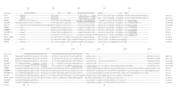
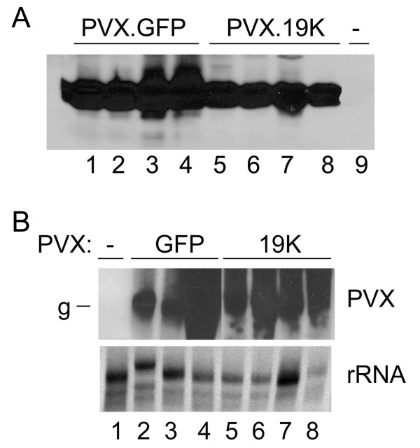
Amino acid sequence analyses indicate that the Soilborne wheat mosaic virus (SBWMV) 19K protein is a cysteine-rich protein (CRP) and shares sequence homology with CRPs derived from furo-, hordei-, peclu- and tobraviruses. Since the hordei- and pecluvirus CRPs were shown to be pathogenesis factors and/or suppressors of RNA silencing, experiments were conducted to determine if the SBWMV 19K CRP has similar activities. The SBWMV 19K CRP was introduced into the Potato virus X (PVX) viral vector and inoculated to tobacco plants. The SBWMV 19K CRP aggravated PVX-induced symptoms and restored green fluorescent protein (GFP) expression to GFP silenced tissues. These observations indicate that the SBWMV 19K CRP is a pathogenicity determinant and a suppressor of RNA silencing.


Figures

Figure 1
Figure 1
Amino acid sequence alignment of the CRPs encoded by furo-, peclu-, tobra- and hordeiviruses. The positions of amino acids are numbered above the alignment. The secondary structure prediction is shown directly above the alignment. Cys and His residues are bold uppercase letters. The leucines of leucine zippers are in bold face. The placement of residues that differ from Pfam are underlined. Vertical bars at the bottom represent where the Pfam family starts and stops. The genus for each virus is indicated on the right of the sequence. Abbreviations and accession numbers for the 33 aligned viruses are used (those displayed are underlined): LyRSV, Lychnis ringspot virus gi_1107721; CWMV-2, Chinese wheat mosaic virus gi_14270345; CWMV, gi_9635448; OGSV Oat golden stripe virus, gi_9635452; SBWMV-NE88 gi_9632360; SBWMV-NE gi_1449160; SBWMV OKl-1, gi_1085914; SBWMV-NY, gi_21630062; SBCMV-Ozz, Soilborne cereal mosaic virus gi_12053756; SBCMV-Fra, gi_9635249; SBCMV-O, gi_6580881; SBCMV-G, gi_6580877; SBCMV-C, gi_6580873; JSBWMV, Japanese soilborne wheat mosaic virus gi_7634693; SCSV, Sorghum chlorotic spot virus gi_21427644; PSLV, Poa semilatent virus gi_321642; BSMV-PV43, Barley stripe mosaic virus gi_19744921; BSMV-RUS, gi_94465; BSMV-JT, gi_808712; BSMV-ND18, gi_1589671; PCV, Peanut clump virus gi_20178597; IPCV, Indian peanut clump virus gi_30018260; TRV-PpK20, Tobacco rattle virus, gi_20522121; TRV-ORY gi_2852339; TRV-Pp085 gi_42733086; TRV-PSG, gi_112699; TRV-PLB, gi_465018; TRV-CAN, gi_1857116; TRV-FL, gi_3033549; TRV-RSTK, gi_6983830; TRV-TCM, gi_112701; PepRSV, Pepper ringspot virus, gi_20178602; PEBV, Pea early browning virus, gi_9632342.
Figure 2
Figure 2
Plants infected with PVX.GFP or PVX.19K at 21 dpi. (A) N. benthamiana plants infected with PVX.GFP (left) and PVX.19K (right). (B, D) PVX.19K-infected N. benthamiana and N. clevelandii plants, respectively, at 21 dpi show systemic necrosis. (C) PVX.GFP-infected N. clevelandii plants. (E, F) C. quinoa and C. amaranticolor leaves infected with PVX.19K (left both panels) and PVX.GFP (right in both panels).
Figure 3
Figure 3
Immunoblot and northern analyses of the PVX infected N. benthamiana plants. (A) Immunoblot analysis conducted using PVX CP antiserum show similar levels of PVX.GFP virus (lanes 1–4) and PVX.19K virus (lanes 5–8). Lane 9 contains extract of non inoculated plants. (B) Northern analysis of RNA isolated from a healthy plant (lane 1), upper noninoculated leaves of PVX.GFP infected plants (lanes 2 – 4) and upper noninoculated leaves of PVX.19K infected plants (lanes 5 – 8). Blots were probed with a GFP sequence probe. The bottom image is the ethidium bromide stained gel showing ribosomal RNAs. Abbrev.: g, genomic RNA.
Figure 4
Figure 4
Evidence for RNA silencing suppression by the SBWMV 19K CRP. (A) nontransgenic N. benthamiana under a UV lamp exhibits red fluorescence due to chlorophyll. (B) GFP-transgenic N. benthamiana (line 16C) exhibits green fluorescence under a UV lamp. (C) GFP was systemically silenced in the 16C transgenic N. benthamiana following infiltration with Agrobacterium. Here in the upper most leaves GFP silencing is vein centric. Systemic GFP silencing is detected initially within 2 weeks. (D) Within 3 weeks, GFP expression is completely silenced in the upper leaves. (E) GFP silenced plant inoculated with PVX.GUS. Emerging tissues of the infected plant remain silenced. (F, G, and H) GFP expression was observed in the emerging tissues of plants that were inoculated with PVX.19K. (I) Northern analyses of total RNAs from nontransgenic tissues (lanes 1, 2) and GFP transgenic tissues (lanes 4 – 7) probed with a labeled GFP sequence probe. Lane 3 is blank. Lanes under the northern blot show ribosomal RNAs on an ethidium bromide stained gel. (J) Northern analysis of total RNAs from 16C plants infiltrated with Agrobacterium containing GFP constructs and probed with a labeled GFP sequence probe. Lanes 1–4 are RNA samples taken from plants that were also inoculated with PVX.19K. Lanes 5–8 are RNA samples taken from plants inoculated with PVX.GUS. Lanes under the northern blot show ribosomal RNAs.

Similar articles

Cited by

References

    1. Dower SK. Cytokines, virokines and the evolution of immunity. Nat Immunol. 2000;1:367–368. doi: 10.1038/80799. - DOI - PubMed
    1. McFadden G, Lalani A, Everett H, Nash P, Xu X. Virus-encoded receptors for cytokines and chemokines. Semin Cell Dev Biol. 1998;9:359–368. doi: 10.1006/scdb.1998.0245. - DOI - PubMed
    1. Bucher E, Hemmes H, de Haan P, Goldbach R, Prins M. The influenza A virus NS1 protein binds small interfering RNAs and suppresses RNA silencing in plants. J Gen Virol. 2004;85:983–991. doi: 10.1099/vir.0.19734-0. - DOI - PubMed
    1. Li WX, Li H, Lu R, Li F, Dus M, Atkinson P, Brydon EW, Johnson KL, Garcia-Sastre A, Ball LA, Palese P, Ding SW. Interferon antagonist proteins of influenza and vaccinia viruses are suppressors of RNA silencing. Proc Natl Acad Sci U S A. 2004;101:1350–1355. doi: 10.1073/pnas.0308308100. - DOI - PMC - PubMed
    1. Kasschau KD, Carrington JC. A counterdefensive strategy of plant viruses: suppression of posttranscriptional gene silencing. Cell. 1998;95:461–470. doi: 10.1016/S0092-8674(00)81614-1. - DOI - PubMed

Publication types