. 2005 Mar 2;3(1):10.
doi: 10.1186/1479-5876-3-10.

Vaccination of metastatic melanoma patients with autologous dendritic cell (DC) derived-exosomes: results of thefirst phase I clinical trial

Affiliations

Vaccination of metastatic melanoma patients with autologous dendritic cell (DC) derived-exosomes: results of thefirst phase I clinical trial

Bernard Escudier et al. J Transl Med. .

Abstract

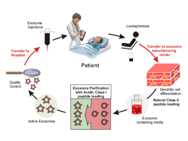
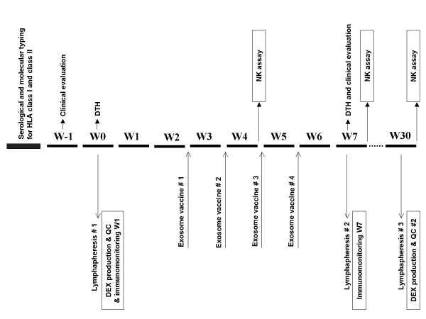
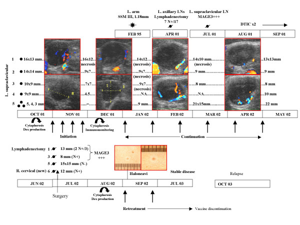
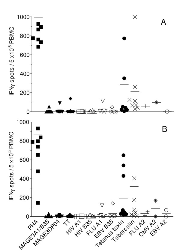
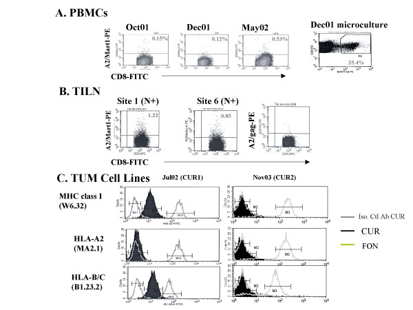
BACKGROUND: DC derived-exosomes are nanomeric vesicles harboring functional MHC/peptide complexes capable of promoting T cell immune responses and tumor rejection. Here we report the feasability and safety of the first Phase I clinical trial using autologous exosomes pulsed with MAGE 3 peptides for the immunization of stage III/IV melanoma patients. Secondary endpoints were the monitoring of T cell responses and the clinical outcome. PATIENTS AND METHODS: Exosomes were purified from day 7 autologous monocyte derived-DC cultures. Fifteen patients fullfilling the inclusion criteria (stage IIIB and IV, HLA-A1+, or -B35+ and HLA-DPO4+ leukocyte phenotype, tumor expressing MAGE3 antigen) were enrolled from 2000 to 2002 and received four exosome vaccinations. Two dose levels of either MHC class II molecules (0.13 versus 0.40 x 10¹⁴ molecules) or peptides (10 versus 100 mug/ml) were tested. Evaluations were performed before and 2 weeks after immunization. A continuation treatment was performed in 4 cases of non progression. RESULTS: The GMP process allowed to harvest about 5 x 10¹⁴ exosomal MHC class II molecules allowing inclusion of all 15 patients. There was no grade II toxicity and the maximal tolerated dose was not achieved. One patient exhibited a partial response according to the RECIST criteria. This HLA-B35+/A2+ patient vaccinated with A1/B35 defined CTL epitopes developed halo of depigmentation around naevi, a MART1-specific HLA-A2 restricted T cell response in the tumor bed associated with progressive loss of HLA-A2 and HLA-BC molecules on tumor cells during therapy with exosomes. In addition, one minor, two stable and one mixed responses were observed in skin and lymph node sites. MAGE3 specific CD4+ and CD8+ T cell responses could not be detected in peripheral blood. CONCLUSION: The first exosome Phase I trial highlighted the feasibility of large scale exosome production and the safety of exosome administration.


Figures

Figure 1
Figure 1
Study design. Different weeks (W) of the study are described. Screening, HLA typing and tumor evaluation were performed within 2 weeks before lymphapheresis. DTH (Multi Mérieux skin test) was performed at the time of lymphapheresis, and exosome vaccine started 3 weeks later.
Figure 2
Figure 2
Schema of exosome purification processes. Exosomes were purified from monocyte derived-DC (MD-DC) culture supernatants according to a good manufacturing process already described [26]. In the second part of the trial, indirect loading of MHC class II peptides (MAGE3247–258.DP04, KKLLTQHFVQENYLEY) was performed on DC cultures followed, after exosome purification by a direct loading of MHC class I (MAGE3168–176.A1/B35; EVDPIGHLY) peptides at 10 or 100 μg/ml at pH 4.2 [22]. Quality control parameters included dosing of exosomal MHC class II molecules, flow cytometry analyses and functional assays using superantigens as described in material and methods.
Figure 3
Figure 3
Clinical outcome of patient #12 during exosome-based vaccination. This patient presented with progressive supraclavicular lymph nodes containing MAGE3 expressing tumor cells in July 2001 when enrolled in the exosomes Phase I trial starting in October 2001. She underwent a first leukapheresis for exosomes production and vaccination (weekly injections in Nov. 2001 during induction therapy and from January 2002 to April 2002 on a three week basis in continuation treatment). The initial size of the target LN are indicated on the left and were followed up by doppler pulsed ultrasonography from Nov 2001 to Dec 2001 (sizes indicated on the right side at the end of the induction therapy). Continuation therapy with exosomes was indicated and maintained clinical stability until the last available exosome dose in Jul. 2002, when she underwent surgery for lymphadenectomy. Results obtained by the pathologists are indicated (N+ if node is invaded by tumor cells, N- if not, MAGE3+ as expression of MAGE3 mRNA in RT-PCR). A second leukapheresis was performed on Aug. 2002 allowing a second therapy with exosomes on a 3 week basis that was continuated until Jul 2004. Six months after exosomes discontinuation (Oct 2004), the patient relapsed in contralateral LN and presented with one lung metastasis.
Figure 4
Figure 4
Evaluation of Tc1/Th1 immune responses to melanoma and viral/recall antigens. A. Before exosome vaccination (W1). B. After exosome inoculation (W7). PBMC obtained at baseline (W1) and after 4 exosome injections are cultured 48 h with the immunizing melanoma antigens i.e Mage 3.A1/B35 or Mage3.DP04 (5 μg/ml) or with viral/recall control antigens (FluMP.A1, HIV.A1, EBV.B35, tetanus anatoxin, tuberculin) or with PHA. The three first patients were also assayed with the universal MHC class II restricted TT peptide. The specific T cell response in each of the evaluated patients is expressed as the number of IFNγ spot forming unit/5 × 105 PBMC.
Figure 5
Figure 5
Antigen spreading and MHC class I loss variant in patient#12. Flow cytometry analyses on serial blood specimen (A) or tumor invaded lymph nodes (B) gating on CD8+ T lymphocytes stained using A2/Mart1 or A2/gag specific fluorescent tetramers. Ex vivo microcultures stimulated with Mart1 peptides and examined according to similar settings. (C) Flow cytometry analyses of two CUR tumor cell lines (pt#12) after the first exosome therapy (continuation treatment) in clinical response and after the second exosome course (second leukapheresis) at relapse for MHC class I (anti-HLA-A2 mAb, anti-HLA-BC mAb and W6.32 Ab) expression. A positive control was included which consisted of a allogeneic HLA-A*0201melanoma line FON.
Figure 6
Figure 6
Lymphocyte recruitment and activation in patient#12's melanoma. T cells that were double-stained by anti-CD3 and anti- CD57 mAb were counted in 2 sections of 6 lymph nodes available from the lymph node dissection specimen obtained before (A. April 2001) and after (B. July 2002) treatment (cf Fig. 3). Counts were performed in 12 sections in total as follows: cells that were double-stained by CD3 and CD57 were counted on the whole section including B cell areas, and the total count was reported to 1 mm2. Results showed an increase of the CD3+CD57+ cells after treatement (mean = 122/mm2) as compared to the count before treatment (mean: 58/mm2).

References

    1. Pardoll DM. Spinning molecular immunology into successful immunotherapy. Nat Rev Immunol. 2002;2:227–238. doi: 10.1038/nri774. - DOI - PubMed
    1. Nestle FO, Banchereau J, Hart D. Dendritic cells: On the move from bench to bedside. Nat Med. 2001;7:761–765. doi: 10.1038/89863. - DOI - PubMed
    1. Steinman RM, Dhodapkar M. Active immunization against cancer with dendritic cells: the near future. Int J Cancer. 2001;94:459–473. doi: 10.1002/ijc.1503. - DOI - PubMed
    1. Dhodapkar MV, Steinman RM, Sapp M, Desai H, Fossella C, Krasovsky J, Donahoe SM, Dunbar PR, Cerundolo V, Nixon DF, Bhardwaj N. Rapid generation of broad T-cell immunity in humans after a single injection of mature dendritic cells. J Clin Invest. 1999;104:173–180. - PMC - PubMed
    1. Schuler-Thurner B, Dieckmann D, Keikavoussi P, Bender A, Maczek C, Jonuleit H, Roder C, Haendle I, Leisgang W, Dunbar R, Cerundolo V, von Den Driesch P, Knop J, Brocker EB, Enk A, Kampgen E, Schuler G. Mage-3 and influenza-matrix peptide-specific cytotoxic T cells are inducible in terminal stage HLA-A2.1+ melanoma patients by mature monocyte-derived dendritic cells. J Immunol. 2000;165:3492–3496. - PubMed