. 2005 Mar 1;33(4):1257-68.
doi: 10.1093/nar/gki272. Print 2005.

The contribution of phosphate-phosphate repulsions to the free energy of DNA bending

Affiliations

The contribution of phosphate-phosphate repulsions to the free energy of DNA bending

Kevin Range et al. Nucleic Acids Res. .

Abstract

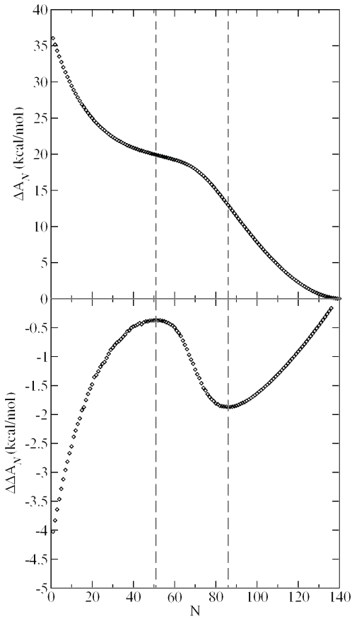
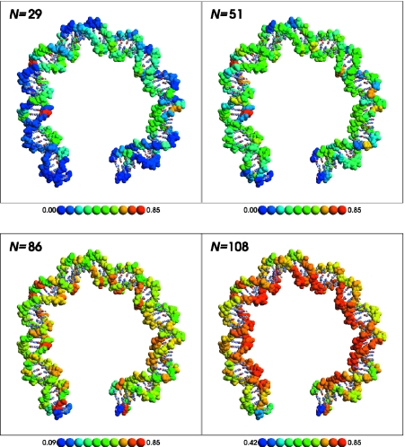
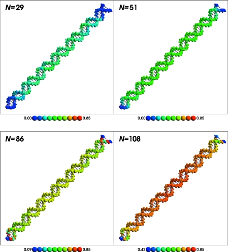
DNA bending is important for the packaging of genetic material, regulation of gene expression and interaction of nucleic acids with proteins. Consequently, it is of considerable interest to quantify the energetic factors that must be overcome to induce bending of DNA, such as base stacking and phosphate-phosphate repulsions. In the present work, the electrostatic contribution of phosphate-phosphate repulsions to the free energy of bending DNA is examined for 71 bp linear and bent-form model structures. The bent DNA model was based on the crystallographic structure of a full turn of DNA in a nucleosome core particle. A Green's function approach based on a linear-scaling smooth conductor-like screening model was applied to ascertain the contribution of individual phosphate-phosphate repulsions and overall electrostatic stabilization in aqueous solution. The effect of charge neutralization by site-bound ions was considered using Monte Carlo simulation to characterize the distribution of ion occupations and contribution of phosphate repulsions to the free energy of bending as a function of counterion load. The calculations predict that the phosphate-phosphate repulsions account for approximately 30% of the total free energy required to bend DNA from canonical linear B-form into the conformation found in the nucleosome core particle.


Figures

Scheme 1
Scheme 1
The 71 bp sequence used to model a full turn of DNA based on the crystallographic structure of a nucleosome core particle (PDB code 1AOI) (50). The human genome consists of ∼3 × 10⁹ bp, two copies of which are contained in the nucleus of each somatic cell. The distance between the base pairs is ∼3.4 Å, leading to a total length of L = 2.04 m. The radius of gyration, including excluded volume effects, can be estimated (1) as Rg1.28×2aL/3, where a is the persistence length, taken here to be 500 Å, leading to a Rg value of 3.34 × 10⁻⁴ m.
Scheme 2
Scheme 2
Thermodynamic cycle for the preferential stabilization of bent versus linear DNA by ions as measured by the change in relative free energy (ΔΔAN) of the electrostatic DNA models. Here, N is the number of site-bound ions and ΔN is the corresponding change in N. Note: ΔΔAN=ΔALΔAB=ΔANΔAN+ΔN.
Figure 1
Figure 1
Top: ΔAN (free energy difference between bent and linear DNA) versus ion load, N, with ε1 = 2 and ε2 = 80. Vertical lines indicate inflection points. Bottom: differential free energy of stabilization, ΔΔAN=ΔABΔAL (Scheme 2) when ΔN = 5 with ε1=2 and ε2=2. Vertical lines indicate extrema.
Figure 2
Figure 2
Average ion occupation for bent DNA at N = 29 (low ion load), 51 [first stationary point of Figure 1 (top) and first inflection point of Figure 1 (bottom)], 86 (second stationary/inflection point), and 108 [ion load predicted by counterion condensation theory (1,12)]. Backbone atoms are shown by a space-filling representation and are colored by their average occupation collected during the course of the Monte Carlo simulation. The average occupation to color mapping is shown in the key below each panel. A movie illustrating how the relative occupation changes as a function of ion load is available in the supplementary information. This Figure was created using RasMol (99).
Figure 3
Figure 3
Average ion occupation for linear DNA at N = 29 (low ion load), 51 [first stationary point of Figure 1 (top) and first inflection point of Figure 1 (bottom)], 86 (second stationary/inflection point) and 108 [ion load predicted by counterion condensation theory (1,12)]. Backbone atoms are shown by a space-filling representation and are colored by their average occupation collected during the course of the Monte Carlo simulation. The average occupation to color mapping is shown in the key below each panel. A movie illustrating how the relative occupation changes as a function of ion load is available in the supplementary information. This Figure was created using RasMol (99).
Figure 4
Figure 4
Effective dielectric constant for solvated phosphate–phosphate repulsions in linear (top) and bent (bottom) DNA. Red points are intra-strand repulsions, blue points are inter-strand repulsions. See Equations 4 and 5 for the definitions of ε′ and R′. Data beyond 30 Å are not shown to better illustrate the deviations at short range; beyond 30 Å the ε′ values are all very close to that of bulk water (ε = 80).

Similar articles

Cited by

References

    1. Bloomfield V.A., Crothers D.M., Tinoco I., Jr . Nucleic Acids: Structures, Properties, and Functions. Sausalito, CA: University Science Books; 2000.
    1. Carr E.A., Mead J., Vershon A.K. α1-induced DNA bending is required for transcriptional activation by the Mcm1-α1 complex. Nucleic Acids Res. 2004;32:2298–2305. - PMC - PubMed
    1. Hardwidge P.R., Zimmerman J.M., Maher L.J., III Charge neutralization and DNA bending by the Escherichia coli catabolite activator protein. Nucleic Acids Res. 2002;30:1879–1885. - PMC - PubMed
    1. Olson W.K., Zhurkin V.B. Modeling DNA deformations. Curr. Opin. Struct. Biol. 2000;10:286–297. - PubMed
    1. Horton N.C., Perona J.J. DNA cleavage by EcoRV endonuclease: two metal ions in three metal ion binding sites. Biochemistry. 2004;43:6841–6857. - PubMed

Publication types