. 2005 Mar 15;118(Pt 6):1321-30.
doi: 10.1242/jcs.01709. Epub 2005 Mar 1.

Higher-order nuclear organization in growth arrest of human mammary epithelial cells: a novel role for telomere-associated protein TIN2

Affiliations

Higher-order nuclear organization in growth arrest of human mammary epithelial cells: a novel role for telomere-associated protein TIN2

Patrick Kaminker et al. J Cell Sci. .

Abstract

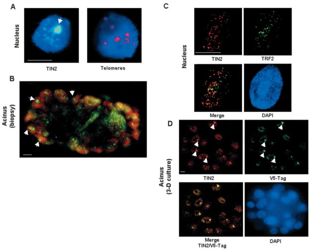
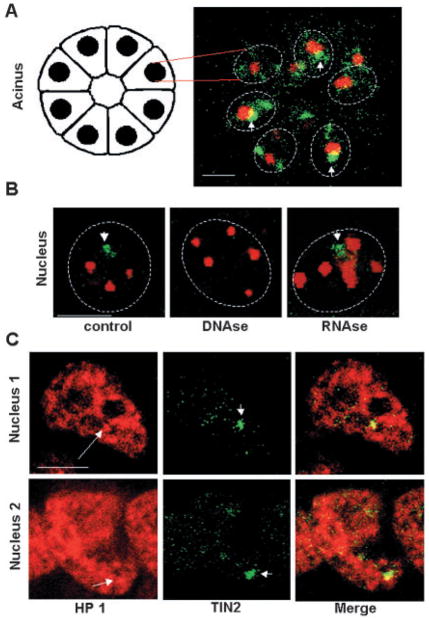
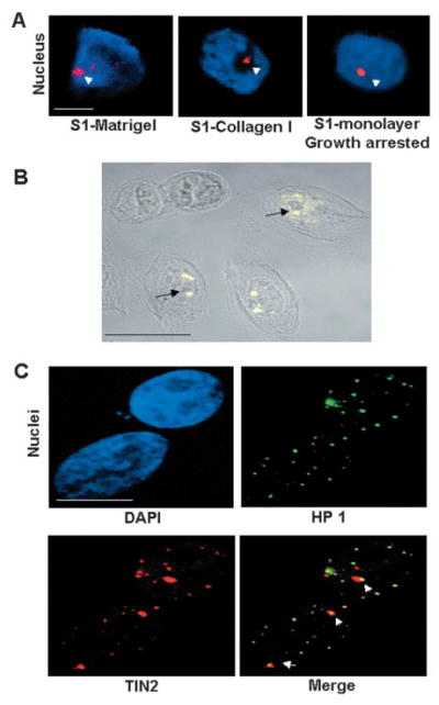
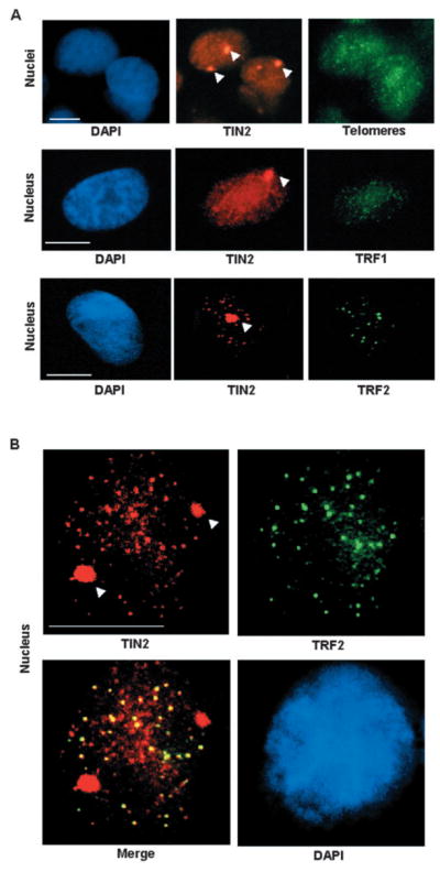
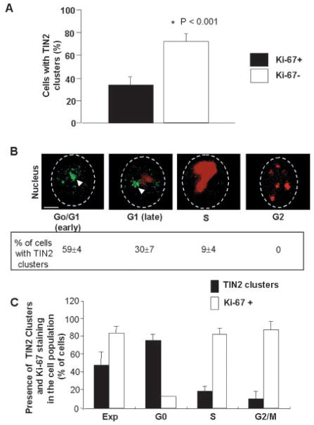
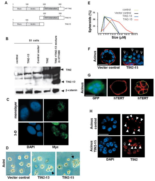
Nuclear organization, such as the formation of specific nuclear subdomains, is generally thought to be involved in the control of cellular phenotype; however, there are relatively few specific examples of how mammalian nuclei organize during radical changes in phenotype, such as those occurring during differentiation and growth arrest. Using human mammary epithelial cells in which growth arrest is essential for morphological differentiation, we show that the arrest of cell proliferation is accompanied by a reorganization of the telomere-associated protein, TIN2, into one to three large nuclear subdomains. The large TIN2 domains do not contain telomeres and occur concomitant with the continued presence of TIN2 at telomeres. The TIN2 domains were sensitive to DNase, but not RNase, occurred frequently, but not exclusively near nucleoli, and overlapped often with dense domains containing heterochromatin protein 1gamma. Expression of truncated forms of TIN2 simultaneously prevented the formation of TIN2 domains and relaxed the stringent morphogenesis-induced growth arrest in human mammary epithelial cells. Here we show that a novel extra-telomeric organization of TIN2 is associated with the control of cell proliferation and identify TIN2 as an important regulator of mammary epithelial differentiation.


Figures

Fig. 1
Fig. 1
Large TIN2 domains are present in HMECs organized into acini. (A) Immunostaining for TIN2 (green) and PNA FISH for telomeres (red) in S1 acini in 3D culture. (B) Immunostaining for TIN2 (green) in a biopsy from normal breast tissue. (C) Dual immunostaining for TIN2 (red) and TRF2 (green) in the nuclei of proliferating 184 HMECs. Colocalization of TIN2 and TRF2 appears yellow. (D) Dual immunostaining for TIN2 (red) and V5 (green) in the nuclei of acini formed by 184 HMECs expressing V5-tagged TIN2 in 3D culture. Nuclei are counterstained with DAPI (blue) in A, C and D, and propidium iodide (red) in B. Images in B and D are confocal sections of acini containing several nuclei. Arrowheads indicate large TIN2 domains. Bar, 5 μm.
Fig. 2
Fig. 2
Large TIN2 domains are often perinucleolar and colocalized with dense HP 1γ foci. (A) Dual immunostaining for TIN2 (green) and nucleolin (red) in S1 acini in 3D culture. The image is a confocal section of an acinus containing several nuclei, illustrated by the drawing on the left. (B) Dual staining for TIN2 (green) and PML (red) in S1 acini. 3D cultures were untreated (control) or treated for 30 minutes with DNase I (DNase) or RNase A (RNase) prior to immunostaining. (C) Dual immunostaining for HP 1γ (red) and TIN2 (green) in S1 acini. Arrows indicate HP 1γ domains that overlap with large TIN2 domains, arrowheads indicate large TIN2 domains and dashed lines delineate the nuclear periphery. Bar, 5 μm.
Fig. 3
Fig. 3
Formation of large TIN2 domains in growth-arrested cells is independent of the differentiation status. (A) Large TIN2 domains (arrowheads) revealed by immunostaining (red) in correctly polarized S1 cells cultured in 3D laminin-rich ECM (S1-Matrigel), S1 cells cultured in 3D collagen I (S1-Collagen I) that display altered polarity, and growth-arrested (EGF-deprived) S1 cells cultured as a monolayer on plastic. Nuclei are counterstained with DAPI (blue). (B) Immunostaining for TIN2 (yellow) in growth-arrested 184 cells cultured as a monolayer on plastic. The superimposed phase-contrast image shows the nucleoli as dark gray subnuclear structures. Arrows indicate large TIN2 domains located next to nucleoli. (C) Dual immunostaining for HP 1γ (green) and TIN2 (red) in growth-arrested 184 cells cultured as a monolayer on plastic. Arrowheads indicate overlapping (yellow) HP 1 staining and large TIN2 domains. Nuclei were counterstained with DAPI (blue). Bar, 5 μm.
Fig. 4
Fig. 4
Formation of large TIN2 domains is independent of telomeres and TRF proteins. (A) HMECs (strain 184) were cultured as a monolayer on plastic and growth-arrested before fixation and dual staining for TIN2 (red) and telomeres (PNA FISH; green) (upper panels), TIN2 (red) and TRF1 (green) (middle panels), and TIN2 (red) and TRF2 (green) (lower panels). (B) Higher magnification images of dual staining for TIN2 (red) and TRF2 (green) showing colocalization at small foci (yellow), indicating telomeric localization. Nuclei were counterstained with DAPI (blue). Arrowheads indicate large TIN2 domains. Bar, 5 μm.
Fig. 5
Fig. 5
Formation of large TIN2 domains correlates with exit from the cell cycle. (A) Percentage of S1 cells with large TIN2 domain(s) and Ki-67 staining. S1 cells were cultured in 3D for 3 days to obtain a mixed population of cycling Ki-67 positive (Ki-67+) and growth-arrested Ki-67 negative (Ki-67−) cells, and then fixed and dual immunostained for TIN2 and Ki-67. Shown is the percentage of cells containing large TIN2 domains (TIN2 clusters) and Ki-67 positive (filled bars) or negative (open bars) staining. Error bars show the s.e.m. for three different experiments. *P<0.001 when compared with Ki-67+ cells. (B) Dual immunostaining for TIN2 (green) and Ki-67 (red) in S1 cells after 3D culture for 3 days. The different phases of the cell cycle were identified by the pattern of Ki-67 staining. The percentage of cells showing large TIN2 domains (TIN2 clusters) in each phase of the cell cycle is given below each panel, and is the mean±s.e.m. of three different experiments. Arrowheads indicate large TIN2 domains and dashed lines delineate the nuclear periphery. (C) Histogram of the percentage of synchronized 184 HMECs with large TIN2 domains (TIN2 clusters) as a function of the cell cycle. Nuclei showing large TIN2 domains were counted as a percentage of total nuclei (revealed by DAPI counterstaining) during exponential (EXP), G0, S and G2-M phases of the cell cycle. Percentages of Ki-67-positive nuclei in each phase are shown. Bar, 2.5 μm.
Fig. 6
Fig. 6
TIN2 controls growth arrest in mammary epithelial cells. (A) Schematic of wild-type TIN2 (WT TIN2), and N-terminally (TIN2-13) and C-terminally (TIN2-15) truncated forms of TIN2. (B) Expression of TIN2 and TIN2-13 in control and TIN2-13 expressing cells shown on western blots. Lanes contained cells used for infection (control), cells expressing TIN2-13 (TIN2-13), cells infected with empty vector (control vector), cells overexpressing wild-type TIN2 (TIN2), control HT1080 fibroblasts expressing exogenous TIN2 and TIN2-13 (TIN2 +TIN2− 13 mixture in HT1080). Arrows indicate the location of the respective bands for TIN2 and TIN2-13. β-catenin was used as a loading control. (C) TIN2-15 expression in monolayer and 3D culture shown by anti-myc immunostaining (green). Nuclei were counterstained with DAPI. Magnification ×1200. (D) Vector control, TIN2-13 and TIN2-15 infected S1 cells were cultured in 3D for 10 days. Shown are phase-contrast images of acini formed by vector control S1 cells and abnormal looking colonies formed by TIN2-13 and TIN2-15 S1 cells. The arrows indicate enlarged and/or irregular multicellular structures. (E) Non-infected S1 cells (control) and vector control, TIN2-13, or TIN2-15 infected S1 cells were cultured in 3D for 10 days. Acini were classified according to six diameter ranges (6–15 μm, 16–25 μm, 26–35 μm, 36–45 μm, 46–55 μm, 56–65 μm). Shown is the percentage of acini in each diameter range out of a total of 400 acini observed in each independent experiment. Three experiments were performed. (F) Immunostaining for the endogenous basement membrane component collagen IV (red) and Ki-67 (green) in vector control or TIN2-15 S1 cells cultured in 3D for 10 days. When proper morphogenesis occurs, acini are surrounded by a continuous basement membrane and >90% of the cells arrest proliferation. One nucleus positive for Ki-67 is seen out of ten nuclei in vector control; five nuclei positive for Ki-67 are seen out of 14 nuclei in TIN2-15. Arrows indicate the absence of collagen IV around part of the TIN2-15 colony. (G) GFP-S1 cells organized in an acinus (left panel). Immunostaining for the endogenous basement membrane component collagen IV (red) (central panel) and α6-integrin (green) and β-catenin (red) (right panel) in hTERT-expressing S1 cells cultured in 3D for 10 days. When proper morphogenesis occurs, in addition to the continuous basement membrane, acini display the localization of α6-integrin at the basal cell membrane (against the basement membrane) and β-catenin at cell-cell junctions. (H) Immunostaining for TIN2 (red) in control or TIN2-15 S1 cells. In the control acinus, eight of nine nuclei, identified by DAPI staining, have a large TIN2 domain (arrowheads). In the acinar structure formed by TIN2-15-expressing cells, four of thirteen nuclei show one or two large TIN2 domains (arrowheads) and one nucleus shows completely fragmented TIN2 domains (arrow). Bar, 25 μm.

Similar articles

Cited by

References

    1. Barbie DA, Kudlow BA, Frock R, Zhao J, Johnson BR, Dyson N, Harlow E, Kennedy BK. Nuclear reorganization of mammalian DNA synthesis prior to cell cycle exit. Mol Cell Biol. 2004;24:595–607. - PMC - PubMed
    1. Bilaud T, Brun C, Ancelin K, Koering CE, Laroche T, Gilson E. Telomeric localization of TRF2, a novel human telobox protein. Nat Genet. 1997;17:236–239. - PubMed
    1. Bissell MJ, Rizki A, Mian IS. Tissue architecture: the ultimate regulator of breast epithelial function. Curr Opin Cell Biol. 2003;6:753–762. - PMC - PubMed
    1. Borden KL. Pondering the promyelocytic leukemia protein (PML) puzzle: possible functions for PML nuclear bodies. Mol Cell Biol. 2002;22:5259–5269. - PMC - PubMed
    1. Braun N, Papadopoulos T, Muller-Hermelink HK. Cell cycle dependent distribution of the proliferation-associated Ki-67 antigen in human embryonic lung cells. Virchows Arch B Cell Pathol Incl Mol Pathol. 1988;56:25–33. - PubMed

Publication types

MeSH terms