. 2005 Mar 1;19(5):530-5.
doi: 10.1101/gad.317405.

Inactivation of TGFbeta signaling in neural crest stem cells leads to multiple defects reminiscent of DiGeorge syndrome

Affiliations

Inactivation of TGFbeta signaling in neural crest stem cells leads to multiple defects reminiscent of DiGeorge syndrome

Heiko Wurdak et al. Genes Dev. .

Abstract

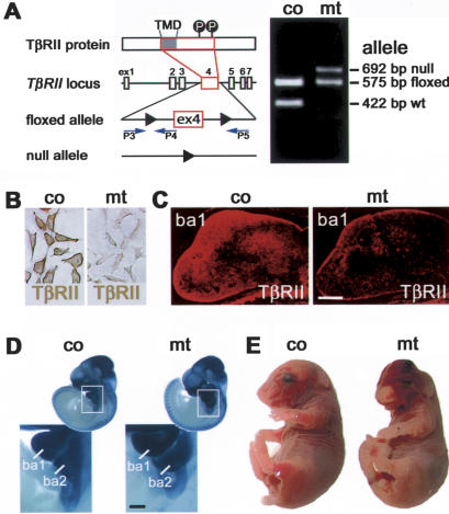
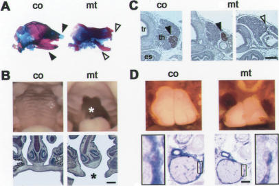
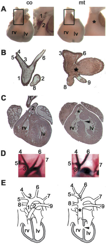
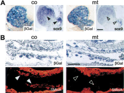
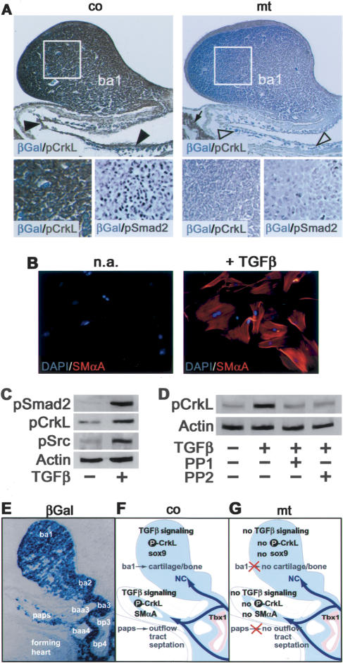
Specific inactivation of TGFbeta signaling in neural crest stem cells (NCSCs) results in cardiovascular defects and thymic, parathyroid, and craniofacial anomalies. All these malformations characterize DiGeorge syndrome, the most common microdeletion syndrome in humans. Consistent with a role of TGFbeta in promoting non-neural lineages in NCSCs, mutant neural crest cells migrate into the pharyngeal apparatus but are unable to acquire non-neural cell fates. Moreover, in neural crest cells, TGFbeta signaling is both sufficient and required for phosphorylation of CrkL, a signal adaptor protein implicated in the development of DiGeorge syndrome. Thus, TGFbeta signal modulation in neural crest differentiation might play a crucial role in the etiology of DiGeorge syndrome.


Figures

Figure 1.
Figure 1.
Neural crest-specific conditional ablation of TβRII. (A) Exon 4 (ex4) of the TβRII locus, encoding the transmembrane domain (TMD) and the intracellular phosphorylation sites (P) of the TβRII protein, is flanked by loxP-sites and deleted in NCSCs upon breeding with wnt1-Cre mice. Identification of the floxed TβRII allele in control (co) and of the recombined TβRII-null allele in mutant (mt) animals by PCR with indicated primers P3, P4, and P5 (Leveen et al. 2002). (B,C) Presence and absence of TβRII (brown/red) in primary NCSC explants (B), and in neural crest cells populating the branchial arch 1 (ba1) in control and mutant embryos at E11.5 (C). (D) Normal distribution of βGal-expressing neural crest cells in the pharyngeal apparatus of E10.5 mutant mice, as assessed by in vivo fate mapping. Branchial arch 1 (ba1) and 2 (ba2) in enlarged areas marked by boxes. (E) Overall appearance of control (co) and mutant (mt) mice at E18.5. Note the craniofacial anomalies in the mutant. Bars, 200 μm.
Figure 2.
Figure 2.
Features of DiGeorge syndrome in TβRII mutant mice. (A) Hypoplastic or absent bone (red) and cartilage (blue) structures (open arrowheads) in mutant (mt) mice at E18. (co) Control. (B) Cleft palate () in macroscopic view (top) and on frontal sections (bottom) in mutant (mt) mice at E18. (C) Parathyroid glands stained for parathyroid hormone (brown; arrowhead) were hypoplastic or undetectable in mutant (mt) embryos at E18 (open arrowhead). (tr) Trachea; (es) esophagus; (th) thyroid gland. (D) Hypoplastic thymus in mutant (mt) mice at E18 (top), correlating with fewer cortical βGal-expressing neural crest cells (blue) compared to control (co) at E13.5 (bottom). Bars: B,C, 200 μm; D, 100 μm.
Figure 3.
Figure 3.
Malformations in the heart of TβRII mutant mice. (A) Ventricular heart at E18 with detailed view of the paired outflow tract in control (co) and of the truncus arteriosus () in mutant (mt) mice. (rv) Right ventricle; (lv) left ventricle; (1) aorta; (2) pulmonary trunk. (B) Frontal section of the normal outflow tract and the truncus arteriosus (). (C) Frontal section of the heart in control and mutant with defective septum between right ventricle (rv) and left ventricle (lv; arrowhead). (D) Abnormal branching of the left carotid artery from the brachiocephalic trunk in mutant mice visualized by intracardial ink injection. (E) Schematic view of abnormal branching and heart defects in the mutant compared to wild-type anatomy. The mutant's left carotid artery (6) arises from the brachiocephalic trunk (3), and the pulmonary arteries (8, 9) originate in the truncus arteriosus (). (4) Right carotid artery; (5) right subclavian artery; (7) left subclavian artery.
Figure 4.
Figure 4.
TβRII-mutant neural crest cells fail to acquire non-neural fates. (A) Equal distribution of βGal-expressing neural crest cells (blue) in the first and second branchial arch of control (co) and TβRII-mutant (mt) mice at E10.5. Presence (arrowheads) and absence (open arrowheads) of sox9, required for normal craniofacial bone and cartilage development, in branchial arches. (B) Presence of βGal-expressing neural crest cells (blue) in the developing aortopulmonary septum of mutant embryos at E10.5. Mutant neural crest cells fail to differentiate into smooth muscle α-actin (SMαA)-expressing cells (red) forming the septum. Bars, 100 μM.
Figure 5.
Figure 5.
TGFβ-dependent CrkL phosphorylation and neural crest differentiation. (A) Mutant (mt) neural crest cells expressing βGal (blue) in branchial arch 1 (ba1), branchial arch 2 (Supplementary Fig. 1), and the developing aorto-pulmonary septum (arrowheads) at E10 show strongly reduced phosphorylation of CrkL (pCrkL; brown; gross and detailed view) and lack nuclear phospho-Smad2 (brown; detailed view) compared with control (co). Note comparable staining intensity for phosphorylated CrkL in βGal-negative tissue (arrow) in mutant (mt) and control (co). (B) Immortalized NCSCs (Monc1 cells) express smooth muscle α-actin (SMαA) upon treatment with TGFβ, while untreated (n.a.) cells are SMαA-negative. (C) Western blot analysis reveals increased Smad2, CrkL, and Src phosphorylation by TGFβ in Monc1 cells. (D) CrkL phosphorylation in TGFβ-treated Monc1 cells is reduced to levels of untreated cells in the presence of Src kinase inhibitors PP1 and PP2, respectively. (E) βGal-expressing neural crest cells populate the pharyngeal apparatus (saggital section). (ba1-3) Branchial arches 1-3; (bp3/4) branchial pouches 3/4; (baa3/4) branchial arch arteries 3/4; (paps) prospective aorto-pulmonary septum. (F) Neural crest cells (NC; blue) present in the pharyngeal apparatus require TGFβ-signaling for CrkL phosphorylation and for expression of the non-neural markers sox9 in the first branchial arch and SMαA in the forming septum of the heart outflow tract. (G) Similar to neural crest cells in Crkol-mutant mice, TβRII-deficient neural crest cells migrate normally into the pharyngeal apparatus. Here, the signal adaptor protein CrkL fails to be phosphorylated in response to TGFβ, and mutant neural crest cells fail to express sox9 and SMαA. Tbx1 expression in the branchial pouch endoderm and early pharyngeal arch patterning is not affected. The failure of mutant neural crest cells to acquire non-neural fates in the early pharyngeal apparatus impairs the development of tissues derived from the pharyngeal apparatus and leads to a DiGeorge-like phenotype.

Similar articles

Cited by

References

    1. Berti L., Mittler, G., Przemeck, G.K., Stelzer, G., Gunzler, B., Amati, F., Conti, E., Dallapiccola, B., Hrabe de Angelis, M., Novelli, G., et al. 2001. Isolation and characterization of a novel gene from the DiGeorge chromosomal region that encodes for a mediator subunit. Genomics 74: 320-332. - PubMed
    1. Brault V., Moore, R., Kutsch, S., Ishibashi, M., Rowitch, D.H., McMahon, A.P., Sommer, L., Boussida, O., and Kemler, R. 2001. Inactivation of the β-catenin gene by Wnt1-Cre-mediated deletion results in dramatic brain malformation and failure of craniofacial development. Development 128: 1253-1264. - PubMed
    1. Chen S. and Lechleider, R.J. 2004. Transforming growth factor-β induced differentiation of smooth muscle from a neural crest stem cell line. Circ. Res. 94: 1195-1202. - PubMed
    1. Derynck R. and Zhang, Y.E. 2003. Smad-dependent and Smad-independent pathways in TGF-β family signalling. Nature 425: 577-584. - PubMed
    1. Feller S.M. 2001. Crk family adaptors—Signalling complex formation and biological roles. Oncogene 20: 6348-6371. - PubMed

Publication types

MeSH terms

Substances