. 2005 Mar 1;19(5):603-13.
doi: 10.1101/gad.1273605.

FGF8 initiates inner ear induction in chick and mouse

Affiliations

FGF8 initiates inner ear induction in chick and mouse

Raj K Ladher et al. Genes Dev. .

Abstract

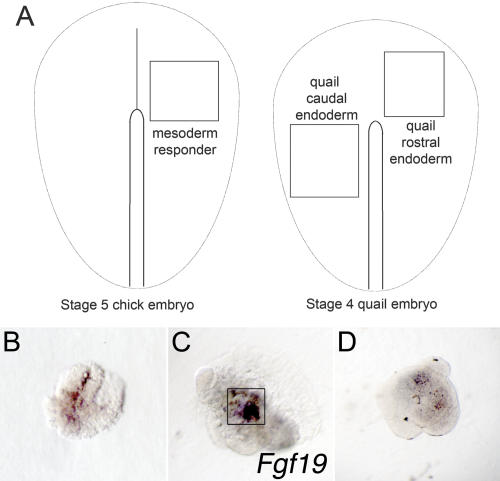
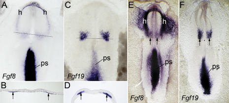
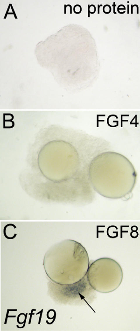
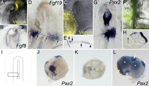
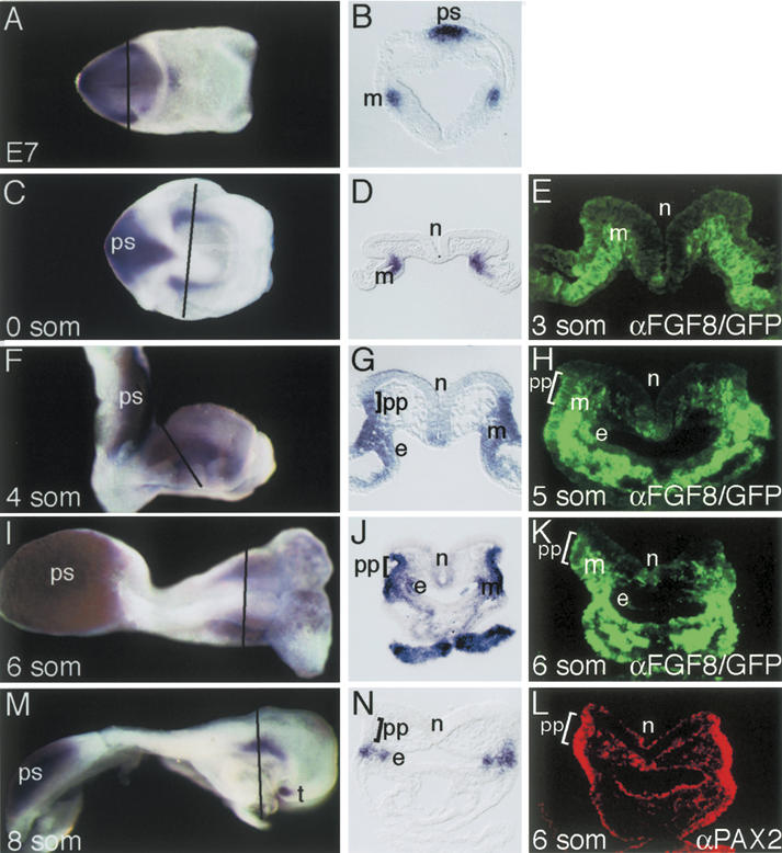
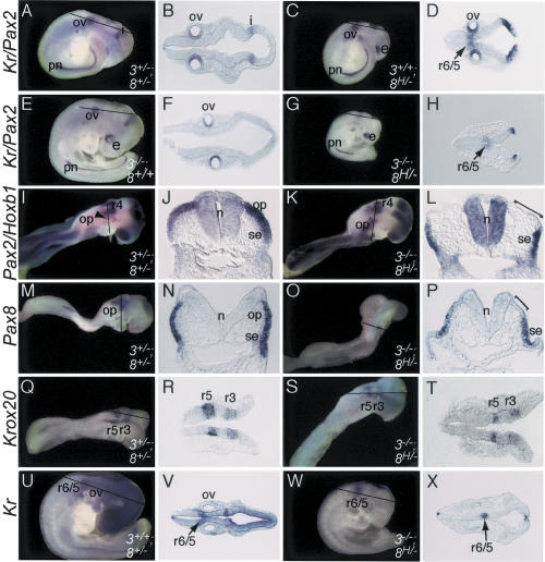
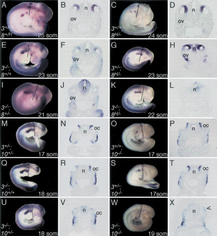
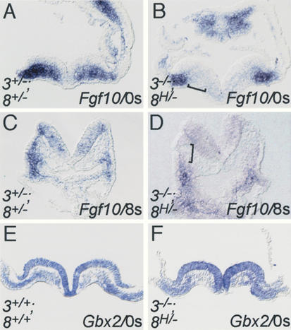
In both chick and mouse, the otic placode, the rudiment of the inner ear, is induced by at least two signals, one from the cephalic paraxial mesoderm and the other from the neural ectoderm. In chick, the mesodermal signal, FGF19, induces neural ectoderm to express additional signals, including WNT8c and FGF3, resulting in induction of the otic placode. In mouse, mesodermal Fgf10 acting redundantly with neural Fgf3 is required for induction of the placode. To determine how the mesodermal inducers of the otic placode are localized, we took advantage of the unique strengths of the two model organisms. We show that endoderm is necessary for otic induction in the chick and that Fgf8, expressed in the chick endoderm subjacent to Fgf19, is both sufficient and necessary for the expression of Fgf19 in the mesoderm. In the mouse, Fgf8 is also expressed in endoderm as well as in other germ layers in the periotic placode region. We show that otic induction fails in embryos null for Fgf3 and hypomorphic for Fgf8 and expression of mesodermal Fgf10 is reduced. Thus, Fgf8 plays a critical upstream role in an FGF signaling cascade required for otic induction in chick and mouse.


Figures

Figure 1.
Figure 1.
Endoderm ablation blocks otic development. The endoderm was removed unilaterally from chick embryos, which were cultured for 24-36 h and then assayed for the otic marker, Pax2.(A) Diagram illustrating endoderm removal (box). (B) Dorsal view showing a smaller otic domain of Pax2 on the operated (left) side. Black line indicates axial level of section in C. (C) Section showing the smaller left otic placode (arrow).
Figure 2.
Figure 2.
Mesodermal Fgf19 expression requires subjacent endoderm. Explanted stage 5 rostral chick mesoderm was cultured alone or with stage 4 quail endoderm. Explants were fixed when staged-matched whole-embryo controls had reached stage 8- and then hybridized with a chick-specific Fgf19 probe. (A) Box in left diagram indicates the mesoderm explanted. Boxes in right diagram indicate the endoderm explanted. (B) Mesodermal explants stripped of ectoderm and endoderm show only background labeling. (C) When recombined with caudal endoderm, mesodermal explants express Fgf19 (boxed). (D) Rostral endoderm fails to induce Fgf19.
Figure 3.
Figure 3.
Chick Fgf8 is expressed in the endoderm underlying mesodermal Fgf19. Chick embryos processed for in situ hybridization with the indicated probes. Lines in A and C indicate the levels of the sections shown in B and D, respectively. (A) Dorsal view of a stage 6 (zero-somite) embryo showing two expression domains of Fgf8 in the cranial endoderm: a horseshoe-shaped area underlying the developing heart rudiments (h) and a transverse domain rostral to the primitive streak (ps). (B) Section showing Fgf8 expression in transverse endodermal domain (arrows). (C) Dorsal view of a stage 6 embryo showing Fgf19 expression in the primitive streak (ps) and in bilateral patches of paraxial mesoderm overlying the Fgf8 transverse-endodermal domain. (D) Section showing bilateral Fgf19 expression in the paraxial mesoderm (arrows). (E) Dorsal view of a stage 8 (three-somite) embryo showing Fgf8 expression in the primitive streak (ps), cranial endodermal domains, and endoderm underlying heart rudiments (h). Note the transverse stripe of endodermal expression (arrows) just rostral to the first pair of somites. (F) Dorsal view of a stage 8 embryo showing Fgf19 expression in the primitive streak (ps) and bilateral paraxial mesodermal patches (arrows) just rostral to the first pair of somites.
Figure 4.
Figure 4.
FGF8 is sufficient for Fgf19 expression. Stripped stage 5 chick mesoderm was cultured in isolation or in the presence of either FGF4 or FGF8. (A) Untreated mesodermal explants fail to express Fgf19 after 8 h of culture. (B) Mesodermal explants treated with FGF4 beads also do not express Fgf19. (C) Mesodermal explants treated with FGF8 beads express Fgf19 (arrow).
Figure 5.
Figure 5.
FGF8 is necessary for Fgf19 expression and inner ear induction. Two plasmids, a Histone-2B/Venus fusion construct and pSilencer-Fgf8, were coelectroporated on one side of stage 4 chick embryos. Treated embryos were cultured for 4-8 or 24-36 h, observed with fluorescent illumination, and then processed for in situ hybridization with probes as indicated. (A) Venus fluorescence (yellow) indicates the electroporated area. (B) Fgf8 transcripts are down-regulated in the region of Venus expression (arrow). (C-E) At stage 7, 4-8 h after electroporation, embryos treated with the pSilencer-Fgf8 vector show a lateral reduction in Fgf19 expression. (C) Only embryos that showed appropriately targeted Venus fluorescence (yellow) were analyzed. (D) Whole mount showing a loss of lateral Fgf19 expression on the electroporated (left) side. Arrows in D and E mark the medial-lateral extent of expression on both the right and left sides. Line marks the level of the section in E.(E) Section showing loss of Fgf19 expression in the lateral paraxial mesoderm on the electroporated (left) side. (F-H) At stages 12-14 (24-36 h after electroporation), embryos treated with the pSilencer-Fgf8 vector show a loss of Pax2 expression and failure of placodal morphology to form on the electroporated (left) side. (F) Only embryos that showed appropriately targeted Venus fluorescence (yellow) were analyzed. (G) Whole mount showing a loss of Pax2 expression on the electroporated side. Line marks the level of the section in H.(H) Section showing loss of Pax2 expression on the electroporated (left) side. In addition, placodal morphology fails to form (arrow), as is evident when compared with the normal Pax2-expressing placode on the right side. (I) Location of three-layered explants (boxed) used in rescue experiments. (J) Control explants express Pax2. (K) Fgf8 siRNA electroporated explants fail to express Pax2.(L) Electroporated explants treated with FGF19 beads (asterisks) express Pax2 (arrows).
Figure 6.
Figure 6.
Mouse Fgf8 is expressed in tissues relevant to early otic development. (A,C,F,I,M) E7.0-E8.0 mouse embryos hybridized with an Fgf8 probe and sectioned in the transverse plane. (B,D,G,J,N) Sections through the otic region (the plane is indicated by a line through each embryo) are shown in the panel to the right of each whole embryo. Whole-mount images are shown in the dorsal (A,C,I) or lateral (F,M) views. Anterior is to the right. (E,H,K) Mouse embryos bearing the Fgf8GFPR allele were sectioned and the FGF8/GFP fusion protein was detected using immunohistochemsitry directed against GFP (green). (L) PAX2 immunohistochemistry (red) was performed on a section adjacent to that shown in K. (A,B) At E7, Fgf8 is expressed in the heart mesoderm (m) and in the primitive streak (ps). (C,D) Fgf8 expression at zero somites in splanchnic mesoderm (m) and primitive streak (ps). (E) At the three-somite stage, the FGF8/GFP fusion protein is localized to splanchnic mesoderm. (F-K) At four to six somites, Fgf8 transcripts and the FGF8/GFP fusion protein are expressed throughout the surface ectoderm, including the prospective placode (pp), pharyngeal endoderm (e), and the splanchnic as well as more dorsal, paraxial, mesoderm (m) and in the primitive streak (ps). At these stages, Fgf8 is also expressed in pharyngeal endoderm (e) and splanchnic as well as more dorsal, paraxial, mesoderm (m) and in the primitive streak (ps). (L) In six-somite embryos, PAX2 expression in the ectoderm of the prospective placode (pp) colocalizes with ectodermal expression of the FGF8/GFP fusion protein in K. (M,N) By eight somites, Fgf8 expression in the surface ectoderm is excluded from the dorsal otic placode (pp) but is maintained in the ventral ectoderm. Expression in the pharyngeal endoderm (e) is maintained. Expression is additionally detected in the telencephalon (t). Expression is absent from hindbrain neural ectoderm (n).
Figure 7.
Figure 7.
Otic, but not hindbrain, development is blocked in Fgf3/Fgf8 double-mutant embryos. (A,C,E,G,I,K,M,O,Q,S,U,W) E8.5-9.5 control and mutant embryos were processed for in situ hybridization with the probes indicated to the left of each row. Embryos were sectioned in either the coronal (B,D,F,H,R,T,V,X) or the transverse plane (J,L,N,P). The section taken through the otic region (the plane is indicated by a line through each embryo) is shown in the panel to the right of each whole embryo. Whole-mount images are shown in the lateral view with rostral to the right. The genotype of each embryo is indicated to the bottom right of each whole-mount panel. (A-H) E9.5 Fgf3/Fgf8 double mutants, but not Fgf3-/- or Fgf8H/- mutants, fail to develop otic vesicles (ov) or express Pax2 in the otic region. The Kr probe also included in this experiment was unsuccessful in labeling the control hindbrain (A,B), so its absence in the Fgf3 mutant (E,F) could not be interpreted. However, the Kr probe did label the Fgf8 mutant (C,D) and double-mutant (G,H) hindbrains, suggesting that r5 and r6 were normal in these embryos. (I-L) In E8.5 control embryos, Pax2 is expressed throughout the otic placode (op) and ventral surface ectoderm (se). In Fgf3/Fgf8 double mutants, Pax2 expression is lost in the dorsal surface ectoderm (indicated by a bracket). Hoxb1 expression in r4 is not affected in Fgf3/Fgf8 double mutants. (M-P) At E8.5, Pax8 expression occurs throughout the otic placode (op) and ventral surface ectoderm (se) of control embryos. In Fgf3/Fgf8 double mutants, Pax8 expression is lost in the dorsal surface ectoderm (indicated by a bracket). (Q-T) Krox20 expression in r3 and r5 is not affected in Fgf3/Fgf8 double mutants. (U-X) Kr expression in r6 and r5 is not affected in Fgf3/Fgf8 double mutants. (pn) Pro-nephros; (i) isthmus; (e) eye; (n) neural ectoderm.
Figure 8.
Figure 8.
In the absence of Fgf3, otic development has a similar dependence on Fgf8 and Fgf10. E9.5 embryos isolated from intercrosses of the Fgf3 and Fgf8 alleles or the Fgf3 and Fgf10 alleles were processed for in situ hybridization with a Gbx2 probe and sectioned in the transverse plane. A section taken through the otic region (the plane is indicated by a line through each embryo) is shown in the panel to the right of each whole embryo. Whole-mount images are labeled with the genotype and somite (som) number and shown in the lateral view with rostral to the right. (A-L) In control, Fgf3+/+;Fgf8H/-, and Fgf3+/-;Fgf8H/- embryos, Gbx2 is expressed normally in the dorsomedial aspect of the otic vesicle (ov). In Fgf3-/-;Fgf8+/+ and Fgf3-/-;Fgf8+/- embryos, Gbx2 expression is diminished and absent, respectively. In Fgf3/Fgf8 double mutants, no otic vesicle and no Gbx2 expression is detected. (M-X) In control, Fgf3+/+;Fgf10-/-, and Fgf3+/-;Fgf10-/- embryos Gbx2 is expressed normally in the dorsomedial region of the otic cup (oc). In the Fgf3-/-;Fgf10+/+ embryo the level of Gbx2 expression is diminished in one otic cup, illustrating the variable expressivity of this phenotype. In Fgf3-/-;Fgf10+/- and Fgf3/Fgf10 double-mutant embryos Gbx2 expression is absent from the otic region. In Fgf3/Fgf8 double mutants (K), this absence of Gbx2 expression is coincident with the complete loss of recognizable otic tissue. However, some disorganised ectodermal tissue (devoid of Gbx2 expression) is seen in the Fgf3-/-;Fgf10-/- embryo (arrowhead). (n) Neural ectoderm.
Figure 9.
Figure 9.
The expression of mesodermal Fgf10 is reduced in Fgf3/Fgf8 double mutants. Preplacodal control (A,C,E) and Fgf3/Fgf8 double-mutant (B,D,F) mouse embryos were hybridized with Fgf10 or Gbx2 as indicated in each panel and sectioned in the transverse plane. (A,B) Sections taken through the otic region of zero somite embryos show a reduction in Fgf10 expression in the medial mesoderm (indicated with a bracket). (C,D) Sections taken through the otic region of eight-somite embryos show a reduction in Fgf10 expression in the mesenchyme underlying the dorsal ectoderm, where the otic placode is expected to form. (E,F) Sections taken through the otic region of zero-somite embryos show that mesodermal Gbx2 expression is un-affected in Fgf3/Fgf8 double-mutant embryos.

Similar articles

Cited by

References

    1. Adamska M., Herbrand, H., Adamski, M., Kruger, M., Braun, T., and Bober, E. 2001. FGFs control the patterning of the inner ear but are not able to induce the full ear program. Mech. Dev. 109: 303-313. - PubMed
    1. Alvarez I.S. and Navascues, J. 1990. Shaping, invagination, and closure of the chick embryo otic vesicle: Scanning electron microscopic and quantitative study. Anat. Rec. 228: 315-326. - PubMed
    1. Alvarez Y., Alonso, M.T., Vendrell, V., Zelarayan, L.C., Chamero, P., Theil, T., Bosl, M.R., Kato, S., Maconochie, M., Riethmacher, D., et al. 2003. Requirements for FGF3 and FGF10 during inner ear formation. Development 130: 6329-6338. - PubMed
    1. Anniko M. and Wikstrom, S.O. 1984. Pattern formation of the otic placode and morphogenesis of the otocyst. Am. J. Otolaryngol. 5: 373-381. - PubMed
    1. Baker C.V. and Bronner-Fraser, M. 2001. Vertebrate cranial placodes I. Embryonic induction. Dev. Biol. 232: 1-61. - PubMed

Publication types

MeSH terms

Substances