Ca2+-induced structural changes in phosphorylase kinase detected by small-angle X-ray scattering
- PMID: 15741333
- PMCID: PMC2253434
- DOI: 10.1110/ps.041124705
Ca2+-induced structural changes in phosphorylase kinase detected by small-angle X-ray scattering
Abstract
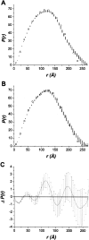
Phosphorylase kinase (PhK), a 1.3-MDa (alphabetagammadelta)(4) hexadecameric complex, is a Ca(2+)-dependent regulatory enzyme in the cascade activation of glycogenolysis. PhK comprises two arched (alphabetagammadelta)(2) octameric lobes that are oriented back-to-back with overall D(2) symmetry and joined by connecting bridges. From chemical cross-linking and electron microscopy, it is known that the binding of Ca(2+) by PhK perturbs the structure of all its subunits and promotes redistribution of density throughout both its lobes and bridges; however, little is known concerning the interrelationship of these effects. To measure structural changes induced by Ca(2+) in the PhK complex in solution, small-angle X-ray scattering was performed on nonactivated and Ca(2+)-activated PhK. Although the overall dimensions of the complex were not affected by Ca(2+), the cation did promote a shift in the distribution of the scattering density within the hydrated volume occupied by the PhK molecule, indicating a Ca(2+)-induced conformational change. Computer-generated models, based on elements of the known structure of PhK from electron microscopy, were constructed to aid in the interpretation of the scattering data. Models containing two ellipsoids and four cylinders to represent, respectively, the lobes and bridges of the PhK complex provided theoretical scattering profiles that accurately fit the experimental data. Structural differences between the models representing the nonactivated and Ca(2+)-activated conformers of PhK are consistent with Ca(2+)-induced conformational changes in both the lobes and the interlobal bridges.
Figures


Similar articles
-
Cryoelectron microscopy reveals new features in the three-dimensional structure of phosphorylase kinase.Protein Sci. 2005 Apr;14(4):914-20. doi: 10.1110/ps.041123905. Epub 2005 Mar 1. Protein Sci. 2005. PMID: 15741332 Free PMC article.
-
Electrostatic changes in phosphorylase kinase induced by its obligatory allosteric activator Ca2+.Protein Sci. 2007 Mar;16(3):517-27. doi: 10.1110/ps.062577507. Protein Sci. 2007. PMID: 17322534 Free PMC article.
-
A Ca(2+)-dependent global conformational change in the 3D structure of phosphorylase kinase obtained from electron microscopy.Structure. 2002 Jan;10(1):23-32. doi: 10.1016/s0969-2126(01)00678-5. Structure. 2002. PMID: 11796107
-
Protein kinase A targeting and activation as seen by small-angle solution scattering.Eur J Cell Biol. 2006 Jul;85(7):655-62. doi: 10.1016/j.ejcb.2006.01.003. Epub 2006 Feb 7. Eur J Cell Biol. 2006. PMID: 16460833 Review.
-
Effect of molecular crowding on the enzymes of glycogenolysis.Biochemistry (Mosc). 2007 Dec;72(13):1478-90. doi: 10.1134/s0006297907130056. Biochemistry (Mosc). 2007. PMID: 18282137 Review.
Cited by
-
Structure and location of the regulatory β subunits in the (αβγδ)4 phosphorylase kinase complex.J Biol Chem. 2012 Oct 26;287(44):36651-61. doi: 10.1074/jbc.M112.412874. Epub 2012 Sep 11. J Biol Chem. 2012. PMID: 22969083 Free PMC article.
-
Probing the MgATP-bound conformation of the nitrogenase Fe protein by solution small-angle X-ray scattering.Biochemistry. 2007 Dec 11;46(49):14058-66. doi: 10.1021/bi700446s. Epub 2007 Nov 15. Biochemistry. 2007. PMID: 18001132 Free PMC article.
-
Cryoelectron microscopy reveals new features in the three-dimensional structure of phosphorylase kinase.Protein Sci. 2005 Apr;14(4):914-20. doi: 10.1110/ps.041123905. Epub 2005 Mar 1. Protein Sci. 2005. PMID: 15741332 Free PMC article.
-
Electrostatic changes in phosphorylase kinase induced by its obligatory allosteric activator Ca2+.Protein Sci. 2007 Mar;16(3):517-27. doi: 10.1110/ps.062577507. Protein Sci. 2007. PMID: 17322534 Free PMC article.
-
Physicochemical changes in phosphorylase kinase induced by its cationic activator Mg(2+).Protein Sci. 2013 Apr;22(4):444-54. doi: 10.1002/pro.2226. Epub 2013 Feb 21. Protein Sci. 2013. PMID: 23359552 Free PMC article.
References
-
- Brostrom, C.O., Hunkeler, F.L., and Krebs, E.G. 1971. The regulation of skeletal muscle phosphorylase kinase by Ca2+. J. Biol. Chem. 246 1961–1967. - PubMed
-
- Brushia, R.J. and Walsh, D.A. 1999. Phosphorylase kinase: The complexity of its regulation is reflected in the complexity of its structure. Front. Biosci. 4 D618–D641. - PubMed
-
- Burger, D., Cox, J.A., Fischer, E.H., and Stein, E.A. 1982. The activation of rabbit skeletal muscle phosphorylase kinase requires the binding of 3 Ca2+ per δ subunit. Biochem. Biophys. Res. Commun. 105 632–638. - PubMed
-
- Cohen, P. 1973. The subunit structure of rabbit-skeletal-muscle phosphorylase kinase, and the molecular basis of its activation reactions. Eur. J. Biochem. 34 1–14. - PubMed
Publication types
MeSH terms
Substances
Grants and funding
LinkOut - more resources
Full Text Sources
Miscellaneous

