

doi: 10.1110/ps.041231005.
Epub 2005 Mar 1.
Distinguishing multiple chemotaxis Y protein conformations with laser-polarized 129Xe NMR
Affiliations
- PMID: 15741343
- PMCID: PMC2253452
- DOI: 10.1110/ps.041231005
Item in Clipboard
Distinguishing multiple chemotaxis Y protein conformations with laser-polarized 129Xe NMR
Protein Sci.
2005 Apr.
Abstract
The chemical shift of the (129)Xe NMR signal has been shown to be extremely sensitive to the local environment around the atom and has been used to follow processes such as ligand binding by bacterial periplasmic binding proteins. Here we show that the (129)Xe shift can sense more subtle changes: magnesium binding, BeF(3)(-) activation, and peptide binding by the Escherichia coli chemotaxis Y protein. (1)H-(15)N correlation spectroscopy and X-ray crystallography were used to identify two xenon-binding cavities in CheY that are primarily responsible for the shift changes. One site is near the active site, and the other is near the peptide binding site.
Figures
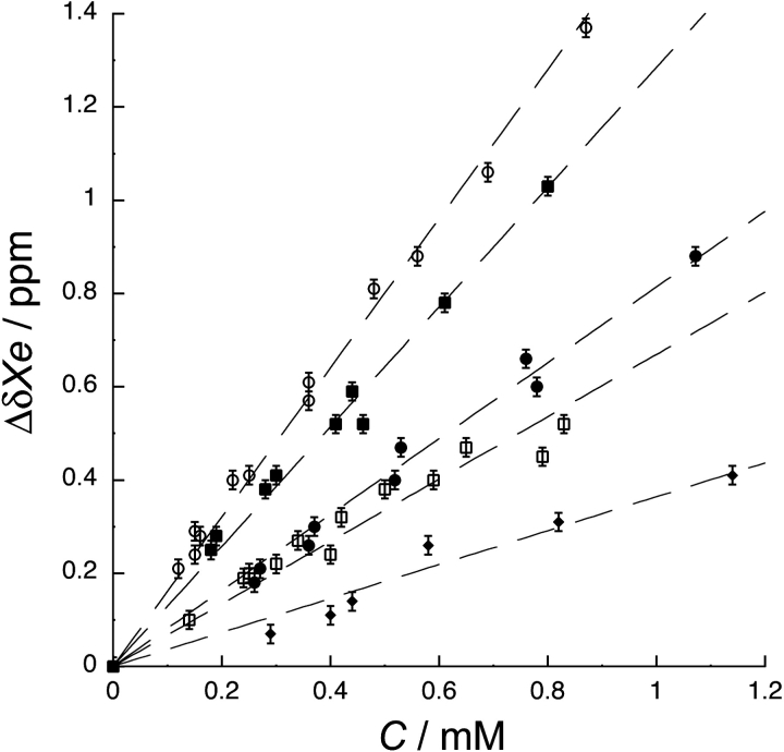
Change in 129Xe chemical shift (ΔδXe) with protein concentration (C) for apo (○), magnesium-bound (▪), activated (•), FliM peptide-bound (□), and denatured (⋄) CheY. Δ δXe is the difference between the 129Xe chemical shift of each titration point and buffer. 129Xe chemical shift values and error bars (± 0.01 ppm) were obtained from peak fits. The concentration-normalized 129Xe chemical shifts, or slopes of the lines, are 1.6 ± 0.1 ppm mM−1, 1.3 ± 0.1 ppm mM−1, 0.8 ± 0.1 ppm mM−1, 0.7 ± 0.1 ppm mM−1, and 0.4 ± 0.1 ppm mM−1, for apo, magnesium-bound, activated, FliM-bound, and denatured CheY, respectively. 129Xe chemical shift is sensitive to four different CheY functional states.
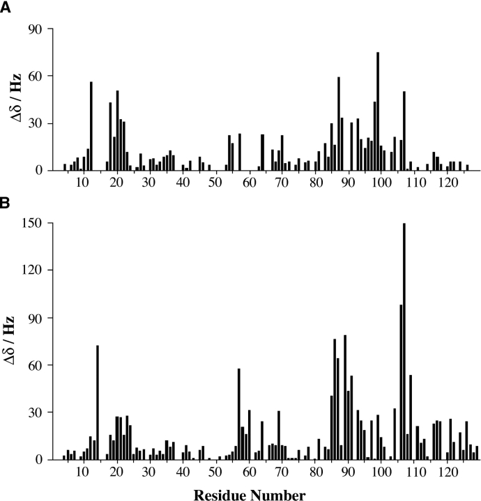
Histogram plots of the total xenon-induced shift for each assigned residue. (A) Shifts induced by 40 mM xenon for apo CheY. (B) Shifts induced by 45 mM xenon for activated CheY. Similar segments of the protein undergo xenon-induced shifts for both CheY conformations.
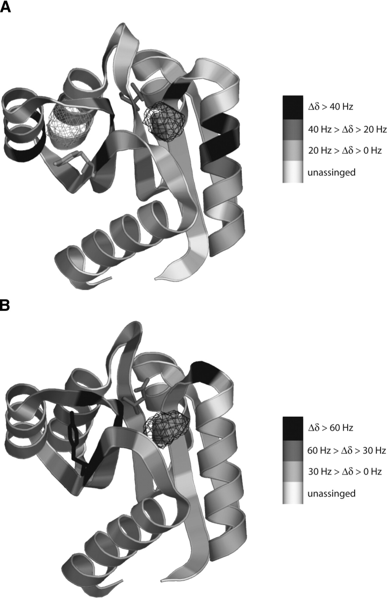
Xenon-induced shifts from Figure 2 ▶ mapped on the backbone of apo CheY (A) (Simonovic and Volz 2001) and activated CheY (B) (this work). Residues are shaded according to the magnitude of their xenon-induced shifts. Unassigned residues are shaded white. For both conformations of CheY shifting residues cluster around two cavities identified by VOIDOO, the H1-β3 cavity (black mesh) and the β4-H4 cavity (gray mesh). Magnesium-binding and BeF3− activation occur in the active site, directly above the H1-β3 cavity. The side chains of Tyr106 and Asp57 are shown in both structures. Activation at Asp57 results in Tyr106 changing its position from “out” to “in,” filling the β4-H4 cavity. Cavities were calculated with VOIDOO (Jones et al. 1991) and images generated with PyMol (DeLano Scientific LLC).
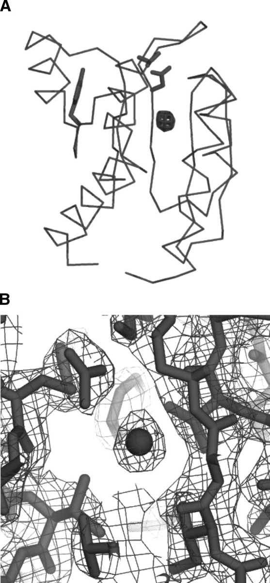
X-ray crystal structure of BeF3−-activated CheY bound to xenon. (A) An Fobs–Fcalc difference electron density omit map (mesh) contoured at 10σ with the xenon atom removed from the model. Electron density from the xenon atom is clearly visible just below the active site inside the H1-β3 cavity. (B) A 2Fobs–Fcalc electron density map (mesh) of the H1-β3 cavity contoured at 1.5σ showing the xenon atom (sphere) and the surrounding residues (sticks).
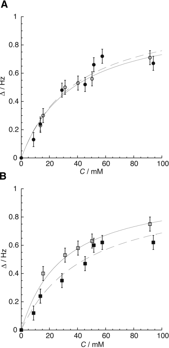
(A) The average of the normalized change in chemical shift (Δ δ) vs. xenon concentration, C, for the amide proton resonances of residues Asp12, Arg18, Ile20, and Asp57, which line the H1-β3 cavity, for the apo (○, solid line) and the activated (•, dashed line) forms of CheY. (B) The average of the normalized change in chemical shift (Δ δ) vs. xenon concentration, C, for the amide proton resonances of residues Tyr106 and Val107, which line the β4-H4 cavity, for the apo (□, solid line) and activated (▪, dashed line) conformations of CheY. Chemical shift changes were normalized according to the limiting shift obtained by an initial fit for each residue. These normalized values were averaged for residues unique to the two cavities to obtain the data and error bars shown in this figure. The respective binding constants obtained from these fits are shown in Table 2. Unlike the H1-β3 cavity, the xenon binding affinity of the β4-H4 cavity changes with activation.
Comment in
-
Detection of multiple protein conformations by laser-polarized xenon.Protein Sci. 2005 Apr;14(4):847. doi: 10.1110/ps.051398705. Protein Sci. 2005. PMID: 15772304 Free PMC article. No abstract available.
Similar articles
-
Detection of multiple protein conformations by laser-polarized xenon.Protein Sci. 2005 Apr;14(4):847. doi: 10.1110/ps.051398705. Protein Sci. 2005. PMID: 15772304 Free PMC article. No abstract available.
-
Structural investigation of pig metmyoglobin by 129Xe NMR spectroscopy.Biochim Biophys Acta. 2004 Sep 24;1674(2):182-92. doi: 10.1016/j.bbagen.2004.06.011. Biochim Biophys Acta. 2004. PMID: 15374622
-
Nuclear magnetic resonance spectroscopy reveals the functional state of the signalling protein CheY in vivo in Escherichia coli.Mol Microbiol. 2003 Sep;49(5):1191-200. doi: 10.1046/j.1365-2958.2003.03628.x. Mol Microbiol. 2003. PMID: 12940980
-
Use of 19F NMR to probe protein structure and conformational changes.Annu Rev Biophys Biomol Struct. 1996;25:163-95. doi: 10.1146/annurev.bb.25.060196.001115. Annu Rev Biophys Biomol Struct. 1996. PMID: 8800468 Free PMC article. Review.
-
Response regulation in bacterial chemotaxis.J Cell Biochem. 1993 Jan;51(1):41-6. doi: 10.1002/jcb.240510109. J Cell Biochem. 1993. PMID: 8381790 Review.
Cited by
-
Insights into correlated motions and long-range interactions in CheY derived from molecular dynamics simulations.Biophys J. 2007 Mar 15;92(6):2062-79. doi: 10.1529/biophysj.106.081950. Epub 2006 Dec 15. Biophys J. 2007. PMID: 17172298 Free PMC article.
-
Xenon-Protein Interactions: Characterization by X-Ray Crystallography and Hyper-CEST NMR.Methods Enzymol. 2018;602:249-272. doi: 10.1016/bs.mie.2018.02.005. Epub 2018 Mar 15. Methods Enzymol. 2018. PMID: 29588032 Free PMC article.
-
NMR Hyperpolarization Techniques of Gases.Chemistry. 2017 Jan 18;23(4):725-751. doi: 10.1002/chem.201603884. Epub 2016 Dec 5. Chemistry. 2017. PMID: 27711999 Free PMC article. Review.
-
Hyperpolarized krypton-83 as a contrast agent for magnetic resonance imaging.Proc Natl Acad Sci U S A. 2005 Dec 20;102(51):18275-9. doi: 10.1073/pnas.0509419102. Epub 2005 Dec 12. Proc Natl Acad Sci U S A. 2005. PMID: 16344474 Free PMC article.
-
Capturing cooperative interactions with the PSI-MI format.Database (Oxford). 2013 Sep 25;2013:bat066. doi: 10.1093/database/bat066. Print 2013. Database (Oxford). 2013. PMID: 24067240 Free PMC article.
References
-
- Bellsolell, L., Prieto, J., Serrano, L., and Coll, M. 1994. Mg2+ binding to the bacterial chemotaxis protein CheY results in large conformational changes involving its functional surface. J. Mol. Biol. 238 489–495. - PubMed
-
- Bolotovsky, R., Steller, I., and Rossman, M.G. 1998. The use of partial reflections for scaling and averaging X-ray area-detector data. J. Appl. Crystallogr. 31 708–717.
-
- Bren, A. and Eisenbach, M. 1998. The N terminus of the flagellar switch protein, FliM, is the binding domain for the chemotactic response regulator, CheY. J. Mol. Biol. 278 507–514. - PubMed
-
- Brünger, A.T., Adams, P.D., Clore, G.M., DeLano, W.L., Gros, P., Grosse-Kunstleve, R.W., Jiang, J.S., Kuszewski, J., Nilges, M., Pannu, N.S., et al. 1998. Crystallography & NMR system: A new software suite for macro-molecular structure determination. Acta Crystallogr. D Biol. Crystallogr. 54 905–921. - PubMed
