. 2005 Apr;14(4):862-72.
doi: 10.1110/ps.041255905. Epub 2005 Mar 1.

Helix periodicity, topology, and dynamics of membrane-associated alpha-synuclein

Affiliations

Helix periodicity, topology, and dynamics of membrane-associated alpha-synuclein

Robert Bussell Jr et al. Protein Sci. 2005 Apr.

Abstract

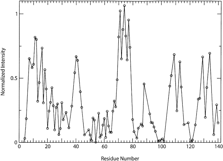
The protein alpha-Synuclein (aS) is a synaptic vesicle-associated regulator of synaptic strength and dopamine homeostasis with a pathological role in Parkinson's disease. The normal function of aS depends on a membrane-associated conformation that is adopted upon binding to negatively charged lipid surfaces. Previously we found that the membrane-binding domain of aS is helical and suggested that it may exhibit an unusual structural periodicity. Here we present a study of the periodicity, topology, and dynamics of detergent micelle-bound aS using paramagnetic spin labels embedded in the micelle or attached to the protein. We show that the helical region of aS completes three full turns every 11 residues, demonstrating the proposed 11/3 periodicity. We also find that the membrane-binding domain is partially buried in the micelle surface and bends toward the hydrophobic interior, but does not traverse the micelle. Deeper submersion of certain regions within the micelle, including the unique lysine-free sixth 11-residue repeat, is observed and may be functionally important. There are no long-range tertiary contacts within this domain, indicating a highly extended configuration. The backbone dynamics of the micelle-bound region are relatively uniform with a slight decrease in flexibility observed toward the C-terminal end. These results clarify the topological features of aS bound to membrane-mimicking detergent micelles, with implications for aS function and pathology.


Figures

Figure 1.
Figure 1.
Effect of the aqueous spin label Mn2+ on NMR backbone resonances of aS. Resonance intensities from a proton–nitrogen correlation spectrum of micelle-bound aS in the presence of 100 μM MnCl2, normalized by resonance intensities in an equivalent spectrum collected in the absence of the spin label, are shown as a function of the corresponding sequence position.
Figure 2.
Figure 2.
Amino acid sequences of human aS and bS. Differences from the aS sequence are indicated in boldface. The imperfect 11-mer repeat regions are delineated by spaces and the core sequence of each repeat is underlined.
Figure 3.
Figure 3.
Correlation of the resonance broadening effects of micelle embedded paramagnetic spin labels 5- and 16-doxylstearate and 4-OH-TEMPO with two different periodicities, 11/3 and 18/5, of micelle-bound aS. The average resonance intensity for all residues at each possible helix projection angle for either an 11/3 (left panels) or 18/5 (right panels) periodicity are shown from spectra of micelle-bound aS in the presence of each of the three spin label reagents. Only residues previously determined to adopt helical structure (1–94) are included. A weaker (stronger) intensity at a given projection angle indicates that residues at this position on the helix are on average closer to (further from) the micelle interior. A sinusoidal pattern is expected for a periodicity that corresponds to that of the actual helical structure of micelle-bound aS.
Figure 4.
Figure 4.
Backbone amide proton chemical shift deviations. The average backbone amide proton chemical shift deviation from the expected random coil value at each possible helix projection angle for either an 11/3 (A) or an 18/5 (B) periodicity are shown. Only residues previously determined to adopt helical structure (1–94) are included. Negative amide proton chemical shift deviations are associated with longer hydrogen bonds and a more hydrophilic environment. Again, a sinusoidal pattern is expected for a periodicity that accurately represents the actual helical structure of the micelle-bound protein.
Figure 5.
Figure 5.
Paramagnetic relaxation enhancement data for three different cysteine mutants of C-terminally truncated detergent micelle-bound aS labeled with the paramagnetic nitroxide spin label MTSL. The intensities of backbone resonances from proton–nitrogen correlation spectra, normalized to those in spectra of spin label-free protein, are displayed as a function of sequence position for each sample. In general, only residues nearby in sequence to the labeling site show significant resonance attenuation, suggesting a lack of long-range tertiary contacts in the protein. The localized attenuation observed around positions 4, 40, and 96 are likely due to nonspecific interactions with free spin label at these positions and are not a result of tertiary interactions.
Figure 6.
Figure 6.
NMR relaxation parameters for backbone 15N nuclei in micelle-bound aS. R1 (longitudinal relaxation rate), R2 (transverse relaxation rate), and the steady-state heteronuclear 1H-15N NOE are plotted as a function of residue number. The NOE data reflect the presence of relatively faster motions, with values near 1 indicating a lack of motion and smaller values indicating increasing mobility. The R2 data reflect slower motions, with higher values of R2 indicating a greater degree of slower vs. faster motions. The high degree of flexibility of the lipid-free C-terminal tail is easily evident in both the NOE and R2 data. The R1 data are relatively less sensitive than the NOE and R2 measurements.

References

    1. Abeliovich, A., Schmitz, Y., Farinas, I., Choi-Lundberg, D., Ho, W.H., Castillo, P.E., Shinsky, N., Verdugo, J.M., Armanini, M., Ryan, A., et al. 2000. Mice lacking α-synuclein display functional deficits in the nigrostriatal dopamine system. Neuron 25 239–252. - PubMed
    1. Ahn, B.H., Rhim, H., Kim, S.Y., Sung, Y.M., Lee, M.Y., Choi, J.Y., Wolozin, B., Chang, J.S., Lee, Y.H., Kwon, T.K., et al. 2002. α-Synuclein interacts with phospholipase D isozymes and inhibits pervanadate-induced phospholipase D activation in human embryonic kidney-293 cells. J. Biol. Chem. 277 12334–12342. - PubMed
    1. Bisaglia, M., Tessari, I., Pinato, L., Bellanda, M., Giraudo, S., Fasano, M., Bergantino, E., Bubacco, L., and Mammi, S. 2005. A topological model of the interaction between α-synuclein and sodium dodecyl sulfate micelles. Biochemistry 44 329–339. - PubMed
    1. Blundell, T., Barlow, D., Borkakoti, N., and Thornton, J. 1983. Solvent-induced distortions and the curvature of α-helices. Nature 306 281–283. - PubMed
    1. Bussell Jr., R. and Eliezer, D. 2001. Residual structure and dynamics in Parkinson’s disease-associated mutants of α-synuclein. J. Biol. Chem. 276 45996–46003. - PubMed

Publication types