

Chemokine receptors in the rheumatoid synovium: upregulation of CXCR5
- PMID: 15743468
- PMCID: PMC1065316
- DOI: 10.1186/ar1475
Chemokine receptors in the rheumatoid synovium: upregulation of CXCR5
Abstract
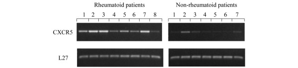
In patients with rheumatoid arthritis (RA), chemokine and chemokine receptor interactions play a central role in the recruitment of leukocytes into inflamed joints. This study was undertaken to characterize the expression of chemokine receptors in the synovial tissue of RA and non-RA patients. RA synovia (n = 8) were obtained from knee joint replacement operations and control non-RA synovia (n = 9) were obtained from arthroscopic knee biopsies sampled from patients with recent meniscal or articular cartilage damage or degeneration. The mRNA expression of chemokine receptors and their ligands was determined using gene microarrays and PCR. The protein expression of these genes was demonstrated by single-label and double-label immunohistochemistry. Microarray analysis showed the mRNA for CXCR5 to be more abundant in RA than non-RA synovial tissue, and of the chemokine receptors studied CXCR5 showed the greatest upregulation. PCR experiments confirmed the differential expression of CXCR5. By immunohistochemistry we were able to detect CXCR5 in all RA and non-RA samples. In the RA samples the presence of CXCR5 was observed on B cells and T cells in the infiltrates but also on macrophages and endothelial cells. In the non-RA samples the presence of CXCR5 was limited to macrophages and endothelial cells. CXCR5 expression in synovial fluid macrophages and peripheral blood monocytes from RA patients was confirmed by PCR. The present study shows that CXCR5 is upregulated in RA synovial tissue and is expressed in a variety of cell types. This receptor may be involved in the recruitment and positioning of B cells, T cells and monocytes/macrophages in the RA synovium. More importantly, the increased level of CXCR5, a homeostatic chemokine receptor, in the RA synovium suggests that non-inflammatory receptor-ligand pairs might play an important role in the pathogenesis of RA.
Figures


Similar articles
-
Differential expression of chemokine receptors on peripheral blood, synovial fluid, and synovial tissue monocytes/macrophages in rheumatoid arthritis.Arthritis Rheum. 2001 May;44(5):1022-32. doi: 10.1002/1529-0131(200105)44:5<1022::AID-ANR181>3.0.CO;2-N. Arthritis Rheum. 2001. PMID: 11352233
-
Monocytes/macrophages express chemokine receptor CCR9 in rheumatoid arthritis and CCL25 stimulates their differentiation.Arthritis Res Ther. 2010;12(4):R161. doi: 10.1186/ar3120. Epub 2010 Aug 25. Arthritis Res Ther. 2010. PMID: 20738854 Free PMC article.
-
Orphan receptor GPR15/BOB is up-regulated in rheumatoid arthritis.Cytokine. 2014 Jun;67(2):53-9. doi: 10.1016/j.cyto.2014.02.015. Epub 2014 Mar 22. Cytokine. 2014. PMID: 24725539 Free PMC article.
-
Monocytes in rheumatoid arthritis: Circulating precursors of macrophages and osteoclasts and, their heterogeneity and plasticity role in RA pathogenesis.Int Immunopharmacol. 2018 Dec;65:348-359. doi: 10.1016/j.intimp.2018.10.016. Epub 2018 Oct 23. Int Immunopharmacol. 2018. PMID: 30366278 Review.
-
[Rheumatoid arthritis: new developments in the pathogenesis with special reference to synovial fibroblasts].Z Rheumatol. 2001 Oct;60(5):309-18. doi: 10.1007/s003930170030. Z Rheumatol. 2001. PMID: 11759230 Review. German.
Cited by
-
Variability in synovial inflammation in rheumatoid arthritis investigated by microarray technology.Arthritis Res Ther. 2006;8(2):R47. doi: 10.1186/ar1903. Epub 2006 Feb 16. Arthritis Res Ther. 2006. PMID: 16507157 Free PMC article.
-
The G Protein-Coupled Receptor (GPR) 15 Counteracts Antibody-Mediated Skin Inflammation.Front Immunol. 2020 Aug 14;11:1858. doi: 10.3389/fimmu.2020.01858. eCollection 2020. Front Immunol. 2020. PMID: 32922401 Free PMC article.
-
Secretion of pro-inflammatory cytokines and chemokines and loss of regulatory signals by fibroblast-like synoviocytes in juvenile idiopathic arthritis.Proteomics Clin Appl. 2017 May;11(5-6):1600088. doi: 10.1002/prca.201600088. Epub 2017 Jan 17. Proteomics Clin Appl. 2017. PMID: 28012239 Free PMC article.
-
CXCL13 antibody for the treatment of autoimmune disorders.BMC Immunol. 2015 Feb 12;16(1):6. doi: 10.1186/s12865-015-0068-1. BMC Immunol. 2015. PMID: 25879435 Free PMC article.
-
B-cell phenotype and IgD-CD27- memory B cells are affected by TNF-inhibitors and tocilizumab treatment in rheumatoid arthritis.PLoS One. 2017 Sep 8;12(9):e0182927. doi: 10.1371/journal.pone.0182927. eCollection 2017. PLoS One. 2017. PMID: 28886017 Free PMC article.
References
-
- Takemura S, Braun A, Crowson C, Kurtin PJ, Cofield RH, O'Fallon WM, Goronzy JJ, Weyand CM. Lymphoid neogenesis in rheumatoid synovitis. J Immunol. 2001;167:1072–1080. - PubMed
Publication types
MeSH terms
Substances
LinkOut - more resources
Full Text Sources
Other Literature Sources
Medical
Molecular Biology Databases
