. 2005;7(2):R291-301.
doi: 10.1186/ar1484. Epub 2005 Jan 11.

The role of regulatory T cells in antigen-induced arthritis: aggravation of arthritis after depletion and amelioration after transfer of CD4+CD25+ T cells

Affiliations

The role of regulatory T cells in antigen-induced arthritis: aggravation of arthritis after depletion and amelioration after transfer of CD4+CD25+ T cells

Oliver Frey et al. Arthritis Res Ther. 2005.

Abstract

It is now generally accepted that CD4+CD25+ Treg cells play a major role in the prevention of autoimmunity and pathological immune responses. Their involvement in the pathogenesis of chronic arthritis is controversial, however, and so we examined their role in experimental antigen-induced arthritis in mice. Depletion of CD25-expressing cells in immunized animals before arthritis induction led to increased cellular and humoral immune responses to the inducing antigen (methylated bovine serum albumin; mBSA) and autoantigens, and to an exacerbation of arthritis, as indicated by clinical (knee joint swelling) and histological scores. Transfer of CD4+CD25+ cells into immunized mice at the time of induction of antigen-induced arthritis decreased the severity of disease but was not able to cure established arthritis. No significant changes in mBSA-specific immune responses were detected. In vivo migration studies showed a preferential accumulation of CD4+CD25+ cells in the inflamed joint as compared with CD4+CD25- cells. These data imply a significant role for CD4+CD25+ Treg cells in the control of chronic arthritis. However, transferred Treg cells appear to be unable to counteract established acute or chronic inflammation. This is of considerable importance for the timing of Treg cell transfer in potential therapeutic applications.


Figures

Figure 1
Figure 1
Depletion of CD25-expressing cells by anti-CD25 treatment. Mice immunized with methylated bovine serum albumin (mBSA) were injected intraperitoneally with 0.5 mg PC61 (anti-CD25) or rat IgG as control 4 and 2 days before arthritis induction. Representative example for flow-cytometric assessment of depletion in spleen cells, using a non-cross-reactive anti-CD25 antibody (7D4) at the time of arthritis induction (day 0).
Figure 2
Figure 2
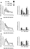
Clinical and histological severity of antigen-induced arthritis (AIA) in CD25-depleted mice. (a) Knee joint swelling (difference in mediolateral joint diameters of arthritic minus nonarthritic knee joints) during the time course of arthritis was higher in CD25-depleted mice. (b) Haematoxylin and eosin stained frontal knee joint sections were scored on a 0–3 point scale at day 14 of AIA for each of the following: severity of synovial hyperplasia, cellular infiltration, cartilage destruction and pannus formation. A score for inflammatory changes (Inf) was calculated by adding the points for synovial hyperplasia and infiltration, and for joint destruction (Dest) by adding the points for cartilage damage and pannus formation. Total arthritis score (Score) was calculated by adding scores for inflammatory changes and joint destruction, giving a maximal AIA score of 12 points. Representative photomicrographs of (c) a control (rat IgG-injected) and (d) a knee joint from an anti-CD25-treated mouse. Ten animals were included in each group in two independent experiments. *P < 0.05, **P < 0.01, ***P < 0.001, versus control.
Figure 3
Figure 3
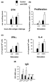
Analysis of in vivo and ex vivo immune responses in CD25-depleted mice. (a) In vivo delayed-type hypersensitivity (DTH) response against methylated bovine serum albumin (mBSA) as a marker for cellular immune response was measured as the increase in ear thickness after intradermal antigen challenge on day 7 of antigen-induced arthritis (AIA). (b) Proliferation, measured as [3H]thymidine incorporation of unstimulated (unst) or mBSA-stimulated (mBSA) draining lymph node cells at day 14 of AIA. (c) Cytokine production was measured with ELISPOT. (d) Serum levels of IgG against mBSA, collagen type I, collagen type II and cartilage proteoglycans were measured using ELISA after 14 days of AIA. Proliferation, DTH reaction and serum IgG titres were tested in 10 animals per group; cytokine production was measured in six animals per group. Data are from one out of two similar experiments. *P < 0.05, **P < 0.01, ***P < 0.001, versus control.
Figure 4
Figure 4
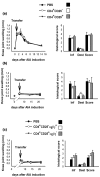
Modulation of antigen-induced arthritis (AIA) by transfer of regulatory T cells (Treg cells). Amelioration of clinical and histological severity of AIA by transfer of 2 × 10⁶ CD4+CD25+ cells freshly isolated from (a) naive or (b) immunized mice at the time of AIA induction (day 0; n = 6 per group). (c) Transfer of 1 × 10⁶ in vitro pre-activated cells at the time of AIA induction (n = 6). #P < 0.05, ##P < 0.01 for CD4+CD25+ versus CD4+CD25-; +P < 0.05, ++P < 0.01 for CD4+CD25+ versus phosphate-buffered saline. Dest, joint destruction; Inf, inflammatory changes; Score; total arthritis score
Figure 5
Figure 5
Transfer of regulatory T cells (Treg cells) cannot cure established arthritis. Pre-activated CD4+CD25+ cells (1 × 10⁶) were transferred on (a) day 1 or (b) day 7 of antigen-induced arthritis (AIA). Arthritis severity was monitored by measurement of knee joint swelling and by histological assessment 14 days after cell transfer (n = 6–7 per group). (c) Also, 1 × 10⁶ pre-activated αEβ7-expressing Treg cells have no curative effect in AIA (n = 8 per group).
Figure 6
Figure 6
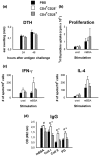
There is no suppression of cellular or humoral methylated bovine serum albumin (mBSA)-specific immunity with transfer of Treg cells. Pre-activated CD4+CD25+ cells (1 × 10⁶) were transferred at the time of antigen-induced arthritis (AIA) induction. (a) Delayed-type hypersensitivity (DTH) reactivity against mBSA in vivo was tested 7 days later by an intradermal antigen challenge into the ears. (b) Antigen-specific proliferation ([3H]thymidine incorporation) and (c) cytokine production (ELISPOT) of draining lymph node cells was measured 14 days after AIA induction. (d) Serum levels of IgG against mBSA, collagen type I, collagen type II and cartilage proteoglycans were measured with ELISA after 14 days of AIA. Proliferation, DTH reaction, cytokine production, and serum IgG titres were tested in six animals per group. #P < 0.05 for CD4+CD25+ versus CD4+CD25-; *P < 0.05 for CD4+CD25- versus phosphate-buffered saline.
Figure 7
Figure 7
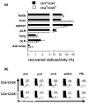
Migration behaviour of regulatory T cells (Treg cells). (a) CD4+CD25+ and CD4+CD25- cells were purified by fluorescence-activated cell sorting (FACS) and labelled with 111In. Cells (1 × 10⁶) were injected intravenously into antigen-induced arthritis (AIA) mice at day 7. After 24 hours radioactivity in isolated organs and the rest of the body was determined using a γ-counter. Thereafter, the total radioactivity recovered per animal was calculated by adding the counts of the organs and the rest of the body. (a) The proportion of radioactivity found in the isolated organs is shown here as a percentage of total recovered radioactivity (n = 6; mean ± standard error of the mean; one representative out of two independent experiments; **P < 0.01). (b) FACS-purified cells were labelled with 5,6-carboxyfluorescein diacetate succinimidyl ester (CFSE) and injected intravenously. After 24 hours single-cell suspensions from draining lymph node (dLN), nondraining peripheral lymph node (pLN), mesenteric lymph node (mLN), spleen and peripheral blood lymphocytes (PBL) were analyzed by FACS. The percentage of CFSE+ cells of the total CD4+ cells was measured. Histogram plots are gated on CD4+ cells after propidium–iodide exclusion of dead cells (n = 3 per group). Higher numbers of CFSEhigh cells are found in the secondary lymphoid organs in the recipients of CD4+CD25- cells.

Similar articles

Cited by

References

    1. Kinne RW, Palombo-Kinne E, Emmrich F. T-cells in the pathogenesis of rheumatoid arthritis villains or accomplices? Biochim Biophys Acta. 1997;1360:109–141. - PubMed
    1. Bluestone JA, Abbas AK. Natural versus adaptive regulatory T cells. Nat Rev Immunol. 2003;3:253–257. doi: 10.1038/nri1032. - DOI - PubMed
    1. Sakaguchi S, Sakaguchi N, Asano M, Itoh M, Toda M. Immunologic self-tolerance maintained by activated T cells expressing IL-2 receptor alpha-chains (CD25). Breakdown of a single mechanism of self-tolerance causes various autoimmune diseases. J Immunol. 1995;155:1151–1164. - PubMed
    1. Stephens LA, Mason D. CD25 is a marker for CD4+ thymocytes that prevent autoimmune diabetes in rats, but peripheral T cells with this function are found in both CD25+ and CD25- subpopulations. J Immunol. 2000;165:3105–3110. - PubMed
    1. Baecher-Allan C, Brown JA, Freeman GJ, Hafler DA. CD4+CD25high regulatory cells in human peripheral blood. J Immunol. 2001;167:1245–1253. - PubMed

Publication types

MeSH terms