Comparative Study
. 2005;7(2):R302-9.
doi: 10.1186/ar1490. Epub 2005 Jan 11.

Autoantibodies specific for apoptotic U1-70K are superior serological markers for mixed connective tissue disease

Affiliations
Comparative Study

Autoantibodies specific for apoptotic U1-70K are superior serological markers for mixed connective tissue disease

Daniëlle Hof et al. Arthritis Res Ther. 2005.

Abstract

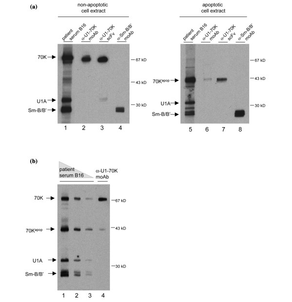
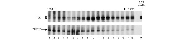
Modifications occurring on autoantigens during cell death have been proposed to have a role in the initiation of autoimmune diseases. Patients suffering from mixed connective tissue disease (MCTD) produce autoantibodies directed to U1 small nuclear ribonucleoprotein (snRNP), and antibodies against a 70 kDa protein component, the U1-70K (70K) protein, are the most prominent. During apoptosis, 70K is cleaved by caspase-3 to a 40 kDa product, which remains associated with the complex. Autoantibodies preferentially recognizing the apoptotic form of 70K have been described previously, and an apoptosis-specific epitope on 70K has been identified. This study shows that 29 of 53 (54%) MCTD sera preferentially recognize the apoptotic form of 70K over intact 70K. Moreover, we show that antibodies directed to an apoptosis-specific epitope on 70K are more specifically associated with MCTD than other anti-70K antibodies, suggesting that apoptotic 70K is a better antigen for the detection of these antibodies in MCTD patients. Longitudinal analysis of 12 MCTD patients showed in several patients that early sera are relatively enriched with antibodies recognizing an apoptosis-specific epitope, and that the levels of these apoptosis-specific antibodies decrease in time. These findings indicate that the early detection of apoptotic 70K is of considerable interest for anti-U1 snRNP-positive patients.


Figures

Figure 1
Figure 1
Anti-U1-70K and anti-70Kapop detection by western blotting. Apoptosis was induced in Jurkat cells by incubation with anisomycin for 8 hours. Western blots were prepared with the resulting cell extracts, and the positions of relevant polypeptides were revealed with patient sera and monoclonal antibodies with the use of a chemiluminescent detection procedure. The positions of the various proteins are indicated on the left, and molecular mass marker positions on the right. (a) U1-70K (70K) detected with a serum from MCTD patient B16 (lanes 1 and 5), anti-70K monoclonal antibody 2.73 (lanes 2 and 6) and an anti-70K single-chain recombinant antibody (scFv; lanes 3 and 7) (70K); lanes 4 and 8, Sm-B/B' detected with a monoclonal anti-Sm-B/B' antibody (ANA125); the position of U1A, which is also recognized by serum from MCTD patient B16 (lanes 1 and 5), was determined by a U1A-specific monoclonal antibody (not shown). In apoptotic cells (lanes 5–8), 70K is present as a 40 kDa species (70Kapop). (b) A serum sample from MCTD patient B16 was applied at 5000-fold (lane 1), 10,000-fold (lane 2) and 20,000-fold (lane 3) dilution on a western blot containing a mixture of non-apoptotic and apoptotic Jurkat cell extracts. In lane 4 the 70K protein was detected with mouse monoclonal antibody 2.73, which reacts much more efficiently with 70K than with 70Kapop.
Figure 2
Figure 2
Longitudinal anti-70K analysis of patient T2. Eighteen serum samples taken over a period of 7 years with approximately equal time intervals were analyzed on western blots containing non-apoptotic and apoptotic Jurkat cell extracts. The positions of U1-70K (70K), of which two isoforms are visible, and 70Kapop are indicated on the left. In lane 19, 70K was detected with mouse monoclonal 2.73, which reacts much more strongly with 70K than with 70Kapop. MoAb, monoclonal antibody.

Similar articles

Cited by

References

    1. Sharp GC, Irvin WS, Tan EM, Gould RG, Holman HR. Mixed connective tissue disease – an apparently distinct rheumatic disease syndrome associated with a specific antibody to an extractable nuclear antigen (ENA) Am J Med. 1972;52:148–159. doi: 10.1016/0002-9343(72)90064-2. - DOI - PubMed
    1. Smolen JS, Steiner G. Mixed connective tissue disease: to be or not to be? Arthritis Rheum. 1998;41:768–777. doi: 10.1002/1529-0131(199805)41:5<768::AID-ART3>3.0.CO;2-Z. - DOI - PubMed
    1. Lerner MR, Steitz JA. Antibodies to small nuclear RNAs complexed with proteins are produced by patients with systemic lupus erythematosus. Proc Natl Acad Sci USA. 1979;76:5495–5499. - PMC - PubMed
    1. Sharp PA. Split genes and RNA splicing. Cell. 1994;77:805–815. doi: 10.1016/0092-8674(94)90130-9. - DOI - PubMed
    1. Lührmann R, Kastner B, Bach M. Structure of spliceosomal snRNPs and their role in pre mRNA splicing. Biochim Biophys Acta. 1990;1087:265–292. - PubMed

MeSH terms