Comparative Study
. 2005;7(2):R380-91.
doi: 10.1186/ar1499. Epub 2005 Jan 26.

Potential involvement of oxidative stress in cartilage senescence and development of osteoarthritis: oxidative stress induces chondrocyte telomere instability and downregulation of chondrocyte function

Affiliations
Comparative Study

Potential involvement of oxidative stress in cartilage senescence and development of osteoarthritis: oxidative stress induces chondrocyte telomere instability and downregulation of chondrocyte function

Kazuo Yudoh et al. Arthritis Res Ther. 2005.

Abstract

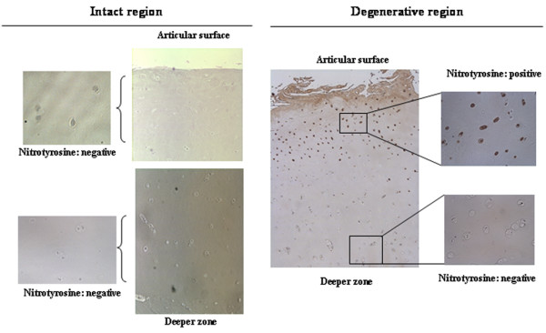
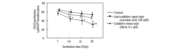
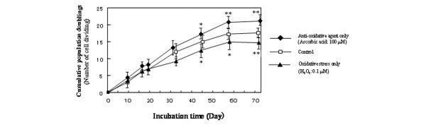
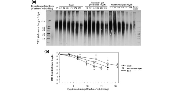
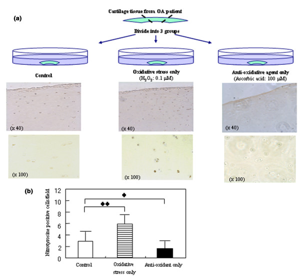
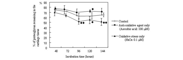
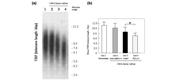
Oxidative stress leads to increased risk for osteoarthritis (OA) but the precise mechanism remains unclear. We undertook this study to clarify the impact of oxidative stress on the progression of OA from the viewpoint of oxygen free radical induced genomic instability, including telomere instability and resulting replicative senescence and dysfunction in human chondrocytes. Human chondrocytes and articular cartilage explants were isolated from knee joints of patients undergoing arthroplastic knee surgery for OA. Oxidative damage and antioxidative capacity in OA cartilage were investigated in donor-matched pairs of intact and degenerated regions of tissue isolated from the same cartilage explants. The results were histologically confirmed by immunohistochemistry for nitrotyrosine, which is considered to be a maker of oxidative damage. Under treatment with reactive oxygen species (ROS; 0.1 micromol/l H2O2) or an antioxidative agent (ascorbic acid: 100.0 micromol/l), cellular replicative potential, telomere instability and production of glycosaminoglycan (GAG) were assessed in cultured chondrocytes. In tissue cultures of articular cartilage explants, the presence of oxidative damage, chondrocyte telomere length and loss of GAG to the medium were analyzed in the presence or absence of ROS or ascorbic acid. Lower antioxidative capacity and stronger staining of nitrotyrosine were observed in the degenerating regions of OA cartilages as compared with the intact regions from same explants. Immunostaining for nitrotyrosine correlated with the severity of histological changes to OA cartilage, suggesting a correlation between oxidative damage and articular cartilage degeneration. During continuous culture of chondrocytes, telomere length, replicative capacity and GAG production were decreased by treatment with ROS. In contrast, treatment with an antioxidative agent resulted in a tendency to elongate telomere length and replicative lifespan in cultured chondrocytes. In tissue cultures of cartilage explants, nitrotyrosine staining, chondrocyte telomere length and GAG remaining in the cartilage tissue were lower in ROS-treated cartilages than in control groups, whereas the antioxidative agent treated group exhibited a tendency to maintain the chondrocyte telomere length and proteoglycan remaining in the cartilage explants, suggesting that oxidative stress induces chondrocyte telomere instability and catabolic changes in cartilage matrix structure and composition. Our findings clearly show that the presence of oxidative stress induces telomere genomic instability, replicative senescence and dysfunction of chondrocytes in OA cartilage, suggesting that oxidative stress, leading to chondrocyte senescence and cartilage ageing, might be responsible for the development of OA. New efforts to prevent the development and progression of OA may include strategies and interventions aimed at reducing oxidative damage in articular cartilage.


Figures

Figure 1
Figure 1
Representative immunohistochemical staining for nitrotyrosine in donor articular cartilage. Cartilage sections were immunostained using an anti-nitrotyrosine antibody. In donor-matched pairs of degenerative and intact regions from same cartilage explants (67-year-old donor), positive immunostaining for nitrotyrosine was observed in chondrocytes and in the cartilage matrix in degenerated regions, whereas the intact region from same cartilage sample showed no positive staining for nitrotyrosine. Original magnification: 40×.
Figure 2
Figure 2
Glycosaminoglycan (GAG) production from cultured chondrocytes under different oxidative conditions. After the incubation times indicated, in the presence of 0.1 μmol/l H2O2 or 100.0 μmol/l ascorbic acid (initial subculture at the start of the experiment: 1 × 10⁵ cells/dish, chondrocytes at passage 2), chondrocytes were collected and transferred to a new culture dish (1 × 10⁵ cells/dish). Following 12 hours of incubation, the amount of GAG in the supernatant was measured using a spectrophotometric assay with dimethylmethylene blue. Values are expressed as the mean ± standard deviation of nine donors (n = 4 culture dishes per treatment group at each incubation period; *P < 0.05, **P < 0.01, versus control group at each incubation time). The H2O2 treated group exhibited a significant decrease in GAG production by chondrocytes as compared with the control group at all incubation times. In the antioxidative agent group the level of proteoglycan production tended to increase as compared with the control group, although no significant differences were observed between the control groups and antioxidative agent groups at any incubation time.
Figure 3
Figure 3
Chondrocyte replicative capacity under the various oxidative conditions. At each subculture (initial subculture at the start of the experiment: 5 × 10⁴ cells/dish, primary culture), the total number of cells in the dish was determined, and the cells (1 × 10⁵ cells/dish) were placed in a new dish. The number of cells that had attached 6 hours after seeding was determined. The increase in cumulative population doublings (number of cell divisions) at each subculture (n = 4 per treatment group) was calculated based on the number of cells attached and the cell yield at the time of the next subcultivation. Cell cultures were considered to have achieved their proliferative limit (senescence) when they did not exceed a twofold increase in 4 weeks. Values are expressed as mean ± standard deviation of four donors. *P < 0.05 and **P < 0.01, versus control group at each incubation time.
Figure 4
Figure 4
Southern blot analysis of chondrocyte telomere lengths in cultured chondrocytes at each passage under the different oxidative conditions. (a) Representative image of Southern blot analysis. Telomere lengths in chondrocytes (1 × 10⁶ cells/dish, initial subculture at the start of the experiment: chondrocytes at passage 3 or 4) were determined using the terminal restriction fragment (TRF) assay. (b) The mean lengths of the chondrocytes were calculated by densitometric molecular weight analysis and were plotted against the number of cell population doublings. *P < 0.05, versus control group at each incubation time. ROS, reactive oxygen species.
Figure 5
Figure 5
Tissue culture of articular cartilage tissue. (a) Representative immunohistochemical staining for nitrotyrosine in cartilage explants treated with reactive oxygen species (ROS) or an antioxidative agent in tissue culture. Osteoarthritis (OA) cartilage explant from a 67-year-old donor was cut and divided into three groups: control group, H2O2 treated group, and antioxidative agent (ascorbic acid-2-O-phosphate [Asc2P]) treated group. After the end of the incubation period (48 hours of incubation), the cartilage sections were immunostained with anti-nitrotyrosine antibody. Original magnifications are given in parentheses. (b) The number of nitrotyrosine positive cells were counted in the 20 areas of tissue-cultured cartilage at 200× magnification (0.785 mm2/field). A statistical analysis of immunostaining was performed. *P < 0.05, **P < 0.01, versus control group.
Figure 6
Figure 6
Glycosaminoglycan (GAG) remaining in the cartilage extract treated with reactive oxygen species (ROS) or antioxidative agent in tissue culture. Catabolic change in articular cartilage matrix was analyzed by determining the GAG content remaining in the cartilage extract relative to the total amount of GAG in the supernatant and the cartilage digest. Values are expressed as mean ± standard deviation of nine donors (three cartilage extracts per donor). *P < 0.05, **P < 0.01, versus control group at each incubation time.
Figure 7
Figure 7
Telomere length of cultured chondrocytes from tissue cultured cartilage explants under the different oxidative conditions. After 144 hours' incubation of tissue culture, chondrocytes were isolated from cartilage explants, which were incubated in the presence or absence of H2O2 (0.1 μmol/l) or ascorbic acid-2-O-phosphate (Asc2P; 100.0 μmol/l). Telomere lengths in chondrocytes (1 × 10⁶chondrocytes of passage 3–4 after isolation) were determined using the terminal restriction fragment (TRF) assay. (a) Representative image of telomere length assay of chondrocytes after 144 hours of incubation. Lane 1, pretreated group (telomere length of isolated chondrocytes from cartilage explants before tissue culture); lane 2, Asc2P + H2O2 treated group; lane 3, control group; lane 4, H2O2 treated group. (b) Treatment with Asc2P (lane 2) showed a tendency to elongate the mean telomere length of chondrocytes in comparison with control. Mean telomere length in H2O2 treated group was significantly shorter than in the control group (n = 9; P < 0.05).

References

    1. Hollander AP, Pidoux I, Reiner A, Rorabeck C, Bourne R, Poole AR. Damage to type II collagen in aging and osteoarthritis starts at the articular surface, originates around chondrocytes, and extends into the cartilage with progressive degeneration. J Clin Invest. 1995;96:2859–2869. - PMC - PubMed
    1. Squires GR, Okouneff A, Ionescu M, Pool AR. The pathobiology of focal lesion development in aging human articular cartilage and molecular matrix changes characteristic of osteoarthritis. Arthritis Rheum. 2003;48:1261–1270. doi: 10.1002/art.10976. - DOI - PubMed
    1. Aurich M, Poole AR, Reiner A, Mollenhauer C, Margulis A, Kuettner KE, Cole AA. Matrix homeostasis in aging normal human ankle cartilage. Arthritis Rheum. 2002;46:2903–2910. doi: 10.1002/art.10611. - DOI - PubMed
    1. Martin JA, Buckwalter JA. Roles of articular cartilage aging and chondrocyte senescence in the pathogenesis of osteoarthritis. Iowa Orthop J. 2001;21:1–7. - PMC - PubMed
    1. Aigner T, Kurz B, Fukui N, Sandell L. Roles of chondrocytes in the pathogenesis of osteoarthritis. Curr Opin Rheumatol. 2002;14:578–584. doi: 10.1097/00002281-200209000-00018. - DOI - PubMed

Publication types

MeSH terms

LinkOut - more resources