Comparative Study
. 2005;7(2):R296-305.
doi: 10.1186/bcr983. Epub 2005 Jan 31.

Claudin-1, -3 and -4 proteins and mRNA expression in benign and malignant breast lesions: a research study

Affiliations
Comparative Study

Claudin-1, -3 and -4 proteins and mRNA expression in benign and malignant breast lesions: a research study

Anna-Mária Tokés et al. Breast Cancer Res. 2005.

Abstract

Introduction: We compared levels of protein and mRNA expression of three members of the claudin (CLDN) family in malignant breast tumours and benign lesions.

Methods: Altogether, 56 sections from 52 surgically resected breast specimens were analyzed for CLDN1, CLDN3 and CLDN4 expression by immunohistochemistry. mRNA was also analyzed using real-time PCR in 17 of the 52 cases.

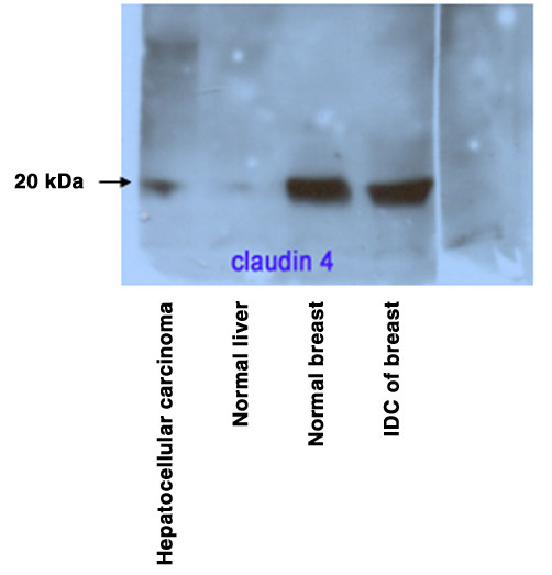
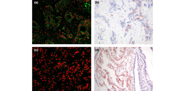
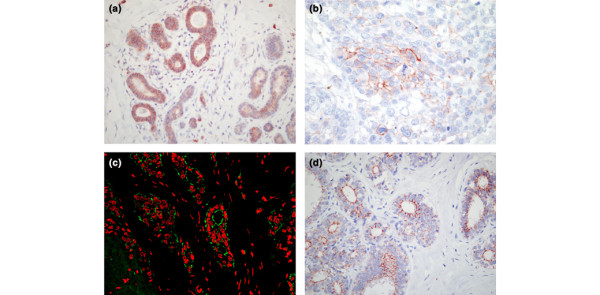
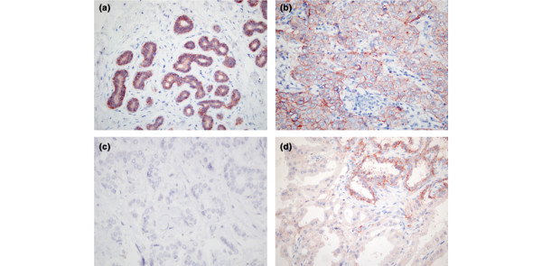
Results: CLDNs were rarely observed exclusively at tight junction structures. CLDN1 was present in the membrane of normal duct cells and in some of the cell membranes from ductal carcinoma in situ, and was frequently observed in eight out of nine areas of apocrine metaplasia, whereas invasive tumours were negative for CLDN1 or it was present in a scattered distribution among such tumour cells (in 36/39 malignant tumours). CLDN3 was present in 49 of the 56 sections and CLDN4 was present in all 56 tissue sections. However, CLDN4 was highly positive in normal epithelial cells and was decreased or absent in 17 out of 21 ductal carcinoma grade 1, in special types of breast carcinoma (mucinous, papillary, tubular) and in areas of apocrine metaplasia. CLDN1 mRNA was downregulated by 12-fold in the sample (tumour) group as compared with the control group using GAPDH as the reference gene. CLDN3 and CLDN4 mRNA exhibited no difference in expression between invasive tumours and surrounding tissue.

Conclusions: The significant loss of CLDN1 protein in breast cancer cells suggests that CLDN1 may play a role in invasion and metastasis. The loss of CLDN4 expression in areas of apocrine metaplasia and in the majority of grade 1 invasive carcinomas also suggests a particular role for this protein in mammary glandular cell differentiation and carcinogenesis.


Figures

Figure 1
Figure 1
The monospecificity of claudin (CLDN)4 antibody using Western blot analysis. CLDN4 protein expression in invasive breast carcinoma, surrounding normal breast and human liver. Western blot analysis was performed on equal amounts of protein from total cell lysates using CLDN4 monoclonal antibody. A single band was shown at approximately 22 kDa for CLDN4. IDC, invasive ductal carcinoma.
Figure 2
Figure 2
Claudin (CLDN)1 positivity in normal breast epithelium, tumour cell membranes and intraductal papilloma. (a) CLDN1 positivity in normal breast epithelium. Normal epithelial cells exhibit intensive CLDN1 positivity in the cell membrane. Image obtained using laser scanning confocal microscopy (MRC 1024; Bio-Rad). Primary antibody was CLDN1 polyclonal antibody (Zymed); secondary antibody was IgG conjugated to Alexafluor 488 (Molecular Probes). Cell nuclei were counter-stained with propidium iodide. Green fluorescence along the cell membranes corresponds to CLDN1; the nuclei are seen in red. (Original magnification: 400×.) (b) CLDN1 positivity in tumour cell membranes. Shown is the immunohistochemical reaction of invasive ductal carcinoma of the breast using polyclonal CLDN-1 antibody. Note the membrane staining only in some scattered tumour cells, as compared with normal epithelial cells (panel a). Sections were counter-stained with Mayer's haemalaun. The chromogen substrate was aminoethylcarbazol. (Original magnification: 600×.) (c) CLDN1 positivity in tumour cell membranes. Shown is the immunohistochemical reaction of invasive ductal carcinoma using polyclonal CLDN1 antibody. There is scattered membrane staining in some tumour cells. The secondary antibody is IgG conjugated to Alexafluor 488. Cell nuclei were counter-stained with propidium iodide. The image was obtained using laser scanning confocal microscopy (MRC 1024; Bio-Rad). (Original magnification: 400×.) (d) CLDN1 positivity in intraductal papilloma. Shown is the immunohistochemical reaction of a breast papilloma with an area of apocrine metaplasia. There is increased CLDN1 positivity in apocrine cells and negative staining in the surrounding papilloma. Sections were counter-stained with Mayer's hemalaun. The chromogen substrate was aminoethylcarbazol. (Original magnification: 200×.)
Figure 3
Figure 3
Claudin (CLDN)3 positivity in benign breast tissue and invasive ductal breast carcinoma. (a) CLDN3 positivity in benign breast tissue. Regular immunohistochemical reaction of benign breast epithelial cells using polyclonal CLDN3 antibody. Continous membrane staining characterizes most of the luminal epithelial cells. Sections were counter-stained with Mayer's haemalaun. The chromogen substrate was aminoethylcarbazol. (Original magnification: 400×.) (b) CLDN3 positivity in invasive ductal breast carcinoma. CLDN3 positivity is apparent in the membranes of some carcinoma cells. Sections were counter-stained with Mayer's hemalaun. The chromogen substrate was aminoethylcarbazol. (Original magnification: 600×.) (c) CLDN3 positivity in benign breast epithelium. CLDN3 positivity is seen in the majority of the membranes of benign epithelial cells. CLDN3 appeared to be localized in normal breast close to the apical end of the cell membranes or in the basolateral membranes of the epithelial cells. The primary antibody was CLDN3 polyclonal antibody (Zymed); the secondary antibody was IgG conjugated to Alexafluor 488 (Molecular Probes). Cell nuclei were counter-stained with propidium iodide. The image was obtained using laser scanning confocal microscopy (MRC 1024; Bio-Rad). (Original magnification: 400×.) (d) CLDN3 positivity in benign breast epithelium. CLDN3 positivity is seen in the membranes of the majority of breast epithelial cells. The positive membrane reaction is incomplete in most of the cells. The primary antibody was CLDN3 polyclonal antibody (Zymed). Sections were counter-stained with Mayer's haemalaun. The chromogen substrate was aminoethylcarbazol. (Original magnification: 400×.)
Figure 4
Figure 4
Claudin (CLDN)4 expression in benign breast epithelium, in invasive ductal carcinomas and in intraductal papilloma. (a) Intense CLDN4 positivity in benign breast epithelium. Shown is the immunohistochemical reaction of benign breast using monoclonal CLDN4 antibody (Zymed). Strong positivity is seen in epithelial cell membranes. Sections were counter-stained with Mayer's haemalaun. The chromogen substrate was aminoethylcarbazol. (Original magnification: 400×.) (b) CLDN4 positivity in invasive ductal breast carcinoma. CLDN4 is expressed in invasive ductal breast carcinoma of grade 3. Positive reaction is evident in the membranes of the tumour cells. Sections were counter-stained with Mayer's haemalaun. The chromogen substrate was aminoethylcarbazol. (Original magnification: 600×.) (c) CLDN4 expression in invasive ductal carcinoma of the breast. Shown is complete loss of CLDN4 expression in invasive ductal breast carcinoma of grade 1 as compared with grade 3 (panel b). Sections were counter-stained with Mayer's haemalaun. The chromogen substrate was aminoethylcarbazol. (Original magnification: 400×.) (d) CLDN4 positivity in intraductal papilloma. Shown is the immunohistochemical reaction of breast papilloma with an area of apocrine metaplasia. The loss of CLDN4 positivity in apocrine cells and the positive staining of other epithelial cells is a 'mirror image' of that seen in the CLDN1 reaction (Fig. 2d). Sections were counter-stained with Mayer's haemalaun. The chromogen substrate was aminoethylcarbazol. (Original magnification: 400×.)

Similar articles

Cited by

References

    1. Gonzalez-Mariscal L, Avila Flores A, Betanzos A. The relationship between structure and function of tight junctions. In: Cereijido M, Anderson JM, editor. Tight Junctions. 2. Boca Raton, FL: CRC Press; 2001. pp. 89–119.
    1. Gonzalez-Mariscal L, Betanzos A, Nava O, Jaramillo BE. Tight junction proteins. Prog Biophys Mol Biol. 2003;81:1–44. doi: 10.1016/S0079-6107(02)00037-8. - DOI - PubMed
    1. Kawabe H, Nakanishi H, Asada M, Fukuhara A, Morimoto K, Takeuchi M, Takai Y. Pilt, a novel peripheral membrane protein at tight junctions in epithelial cells. J Biol Chem. 2001;276:48350–48355. doi: 10.1074/jbc.C100035200. - DOI - PubMed
    1. Katoh M, Katoh M. CLDN23 gene, frequently down-regulated in intestinal-type gastric cancer, is a novel member of CLAUDIN family. Int J Mol Med. 2003;11:683–689. - PubMed
    1. Tsukita S, Furuse M. Occludin and claudins in tight-junction strands: leading or supporting players? Trends Cell Biol. 1999;9:268–273. doi: 10.1016/S0962-8924(99)01578-0. - DOI - PubMed

Publication types

MeSH terms