

BrdU-positive cells in the neonatal mouse hippocampus following hypoxic-ischemic brain injury
- PMID: 15743533
- PMCID: PMC555560
- DOI: 10.1186/1471-2202-6-15
BrdU-positive cells in the neonatal mouse hippocampus following hypoxic-ischemic brain injury
Abstract
Background: Mechanisms that affect recovery from fetal and neonatal hypoxic-ischemic (H-I) brain injury have not been fully elucidated. The incidence of intrapartum asphyxia is approximately 2.5%, but the occurrence of adverse clinical outcome is much lower. One of the factors which may account for this relatively good outcome is the process of neurogenesis, which has been described in adult animals. We used a neonatal mouse model to assess new cells in the hippocampus after H-I injury.
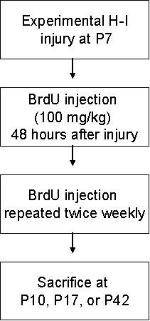
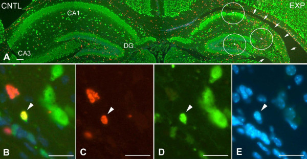
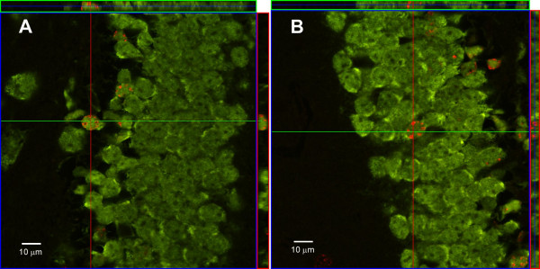
Results: Neonatal mice underwent permanent unilateral carotid ligation on the seventh postnatal day followed by exposure to 8% hypoxia for 75 minutes. The presence of new cells was determined by bromodeoxyuridine (BrdU) incorporation into cells with sacrifice of the animals at intervals. Brain sections were stained for BrdU in combination with neuronal, glial, endothelial and microglial stains. We found a significant increase in BrdU-positive cells in the neonatal mouse hippocampus in the injured area compared to the non-injured area, most prominent in the dentate gyrus (DG) (154.5 +/- 59.6 v. 92.9 +/- 32.7 at 3 days after injury; 68.9 +/- 23.4 v. 52.4 +/- 17.1 at 35 days after injury, p < 0.0011). Among the cells which showed differentiation, those which were stained as either microglial or endothelial cells showed a peak increase at three days after the injury in the DG, injured versus non-injured side (30.5 +/- 17.8 v. 2.7 +/- 2.6, p < 0.0002). As in the adult animal, neurogenesis was significantly increased in the DG with injury (15.0 +/- 4.6 v. 5.2 +/- 1.6 at 35 days after injury, p < 0.0002), and this increase was subsequent to the appearance of the other dividing cells. Numbers of new oligodendrocytes were significantly higher in the DG on the non-injured side (7.0 +/- 24.2 v. 0.1 +/- 0.3, p < 0.0002), suggesting that oligodendrocyte synthesis was reduced in the injured hippocampus.
Conclusion: These findings demonstrate that the neonatal animal responds to brain injury with neurogenesis, much like the adult animal. In addition, H-I insult leads to more neurogenesis than hypoxia alone. This process may play a role in the recovery of the neonatal animal from H-I insult, and if so, enhancement of the process may improve recovery.
Figures
Similar articles
-
Hyperbaric oxygen induces endogenous neural stem cells to proliferate and differentiate in hypoxic-ischemic brain damage in neonatal rats.Undersea Hyperb Med. 2008 Mar-Apr;35(2):113-29. Undersea Hyperb Med. 2008. PMID: 18500076
-
Less neurogenesis and inflammation in the immature than in the juvenile brain after cerebral hypoxia-ischemia.J Cereb Blood Flow Metab. 2007 Apr;27(4):785-94. doi: 10.1038/sj.jcbfm.9600385. Epub 2006 Aug 16. J Cereb Blood Flow Metab. 2007. PMID: 16926844
-
Mesenchymal stem cell treatment after neonatal hypoxic-ischemic brain injury improves behavioral outcome and induces neuronal and oligodendrocyte regeneration.Brain Behav Immun. 2010 Mar;24(3):387-93. doi: 10.1016/j.bbi.2009.10.017. Epub 2009 Oct 31. Brain Behav Immun. 2010. PMID: 19883750
-
Hypoxic-ischemic injury results in acute disruption of myelin gene expression and death of oligodendroglial precursors in neonatal mice.Int J Dev Neurosci. 2001 Apr;19(2):197-208. doi: 10.1016/s0736-5748(00)00075-7. Int J Dev Neurosci. 2001. PMID: 11255033
-
Research advances in neonatal hypoglycemic brain injury.Transl Pediatr. 2012 Oct;1(2):108-15. doi: 10.3978/j.issn.2224-4336.2012.04.06. Transl Pediatr. 2012. PMID: 26835272 Free PMC article. Review. No abstract available.
Cited by
-
Doxycycline inhibits proinflammatory cytokines but not acute cerebral cytogenesis after hypoxia-ischemia in neonatal rats.J Psychiatry Neurosci. 2010 Jan;35(1):20-32. doi: 10.1503/jpn.090061. J Psychiatry Neurosci. 2010. PMID: 20040243 Free PMC article.
-
Functional integration of new neurons into hippocampal networks and poststroke comorbidities following neonatal stroke in mice.Epilepsy Behav. 2010 Aug;18(4):344-57. doi: 10.1016/j.yebeh.2010.05.006. Epub 2010 Jun 17. Epilepsy Behav. 2010. PMID: 20708575 Free PMC article.
-
Pathophysiology of perinatal asphyxia: can we predict and improve individual outcomes?EPMA J. 2011 Jun;2(2):211-30. doi: 10.1007/s13167-011-0100-3. Epub 2011 Jul 26. EPMA J. 2011. PMID: 23199150 Free PMC article.
-
Repeated Ketamine Anesthesia during the Neonatal Period Impairs Hippocampal Neurogenesis and Long-Term Neurocognitive Function by Inhibiting Mfn2-Mediated Mitochondrial Fusion in Neural Stem Cells.Mol Neurobiol. 2024 Aug;61(8):5459-5480. doi: 10.1007/s12035-024-03921-2. Epub 2024 Jan 10. Mol Neurobiol. 2024. PMID: 38200350
-
Sodium Butyrate, a Histone Deacetylase Inhibitor, Exhibits Neuroprotective/Neurogenic Effects in a Rat Model of Neonatal Hypoxia-Ischemia.Mol Neurobiol. 2017 Sep;54(7):5300-5318. doi: 10.1007/s12035-016-0049-2. Epub 2016 Aug 30. Mol Neurobiol. 2017. PMID: 27578020 Free PMC article.
References
Publication types
MeSH terms
Substances
Grants and funding
LinkOut - more resources
Full Text Sources
Other Literature Sources
Research Materials
