. 2005 May 13;280(19):19156-65.
doi: 10.1074/jbc.M502004200. Epub 2005 Mar 1.

Developmental regulation of gonadotropin-releasing hormone gene expression by the MSX and DLX homeodomain protein families

Affiliations

Developmental regulation of gonadotropin-releasing hormone gene expression by the MSX and DLX homeodomain protein families

Marjory L Givens et al. J Biol Chem. .

Abstract

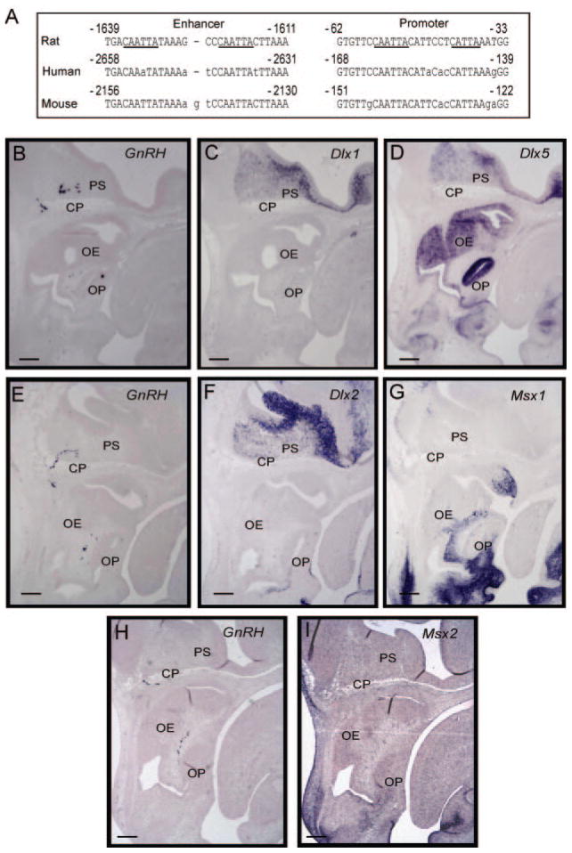
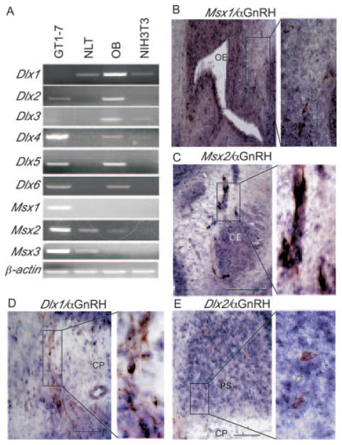
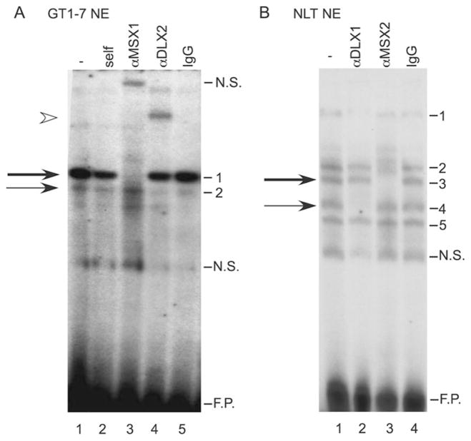
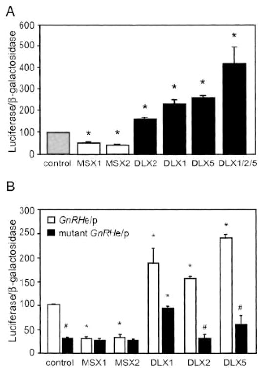
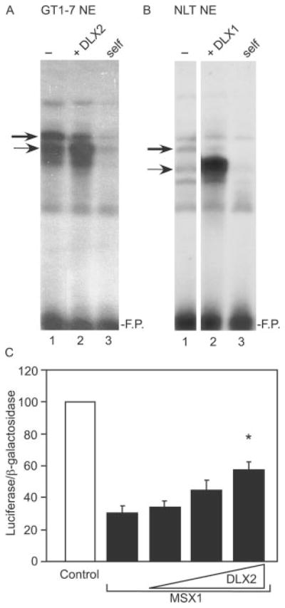
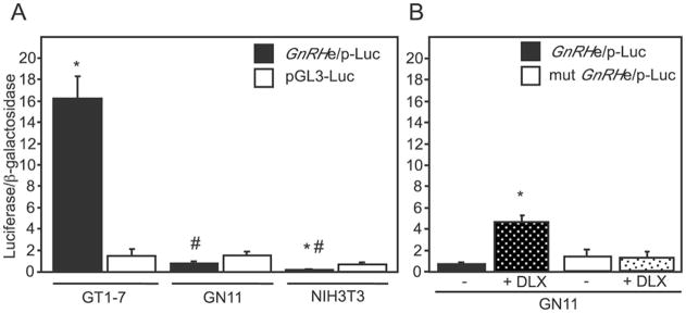
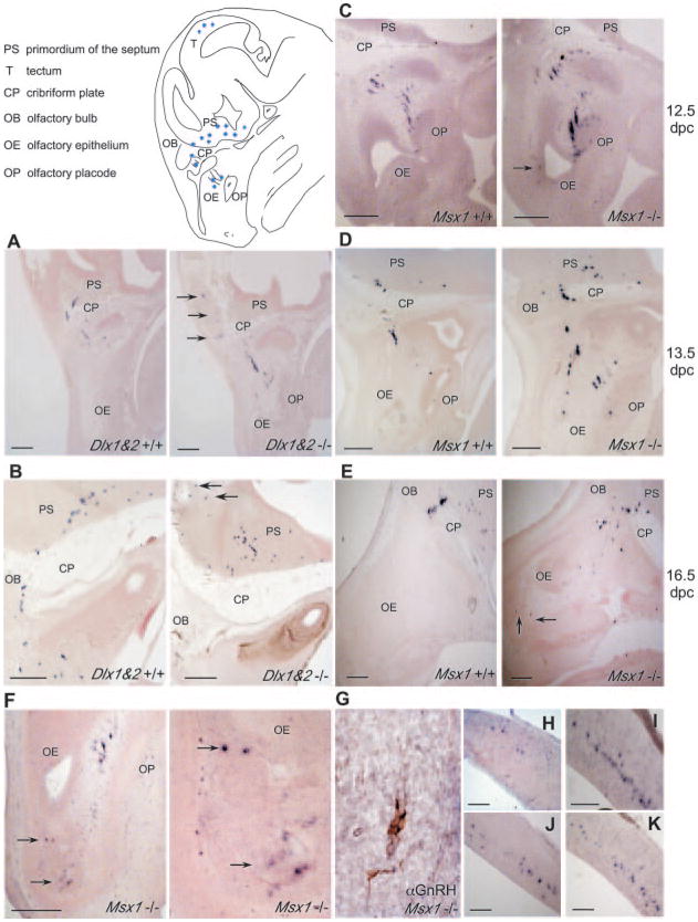
Gonadotropin-releasing hormone (GnRH) is the central regulator of the hypothalamic-pituitary-gonadal axis, controlling sexual maturation and fertility in diverse species from fish to humans. GnRH gene expression is limited to a discrete population of neurons that migrate through the nasal region into the hypothalamus during embryonic development. The GnRH regulatory region contains four conserved homeodomain binding sites (ATTA) that are essential for basal promoter activity and cell-specific expression of the GnRH gene. MSX and DLX are members of the Antennapedia class of non-Hox homeodomain transcription factors that regulate gene expression and influence development of the craniofacial structures and anterior forebrain. Here, we report that expression patterns of the Msx and Dlx families of homeodomain transcription factors largely coincide with the migratory route of GnRH neurons and co-express with GnRH in neurons during embryonic development. In addition, MSX and DLX family members bind directly to the ATTA consensus sequences and regulate transcriptional activity of the GnRH promoter. Finally, mice lacking MSX1 or DLX1 and 2 show altered numbers of GnRH-expressing cells in regions where these factors likely function. These findings strongly support a role for MSX and DLX in contributing to spatiotemporal regulation of GnRH transcription during development.


Figures

Fig. 1
Fig. 1. Conserved homeodomain binding sites in the GnRH upstream sequence and expression of candidate homeodomain regulators
Panel A, four consensus Q50 homeodomain (CAATTA) sites in the rat GnRH gene are located at −1636/−1631, −1623/−1618, −55/−50, and an additional CATTA site in the GnRH promoter at −42/−38 (underlined in the rat sequence). The homologous human and mouse GnRH sequences are shown with lowercase letters indicating mismatches from the rat sequence. B, expression of Msx and Dlx in relation to the migratory GnRH neuron in vivo. In situ hybridization was carried out on parasagittal sections from mouse embryos at 13.5 dpc and probed in series. Sense probes served as controls. Three separate embryos were used for the sections in series, grouped horizontally. Adjacent sections were hybridized using probes for GnRH (panel B), Dlx1 (panel C), and Dlx5 (panel D), or GnRH (panel E), Dlx2 (panel F), and Msx1 (panel G), or GnRH (panel H), and Msx2 (panel I). Abbreviations: olfactory epithelium (OE), cribriform plate (CP), olfactory bulb (OB), primordium of the septum (PS), olfactory placode (OP). Black scale bars represent 100 μm in the lower left of each panel.
Fig. 2
Fig. 2. Expression of Msx and Dlx family members in model GnRH cell lines and co-expression with GnRH in vivo
A, reverse transcriptase PCR analysis was used to identify the Msx and Dlx genes expressed in the GT1-7 and NLT cells. Olfactory bulb (OB) RNA was used as a positive control for the Dlx family members. NIH3T3 RNA is a mouse fibroblast control. β-Actin was used as a control for equal RNA loading. Ethidium-stained, 2% agarose gels are shown here. B–E, Msx and Dlx family members co-express with GnRH. Double in situ hybridization/immunohistochemistry was performed on parasagittal sections of 13.5-dpc embryos with an antibody recognizing GnRH (brown) and RNA probes (blue) for Msx1 (B), Msx2 (C), Dlx1 (D), and Dlx2 (E). The expanded panels to the right indicate higher magnification images of the boxed region to the left. Abbreviations: olfactory epithelium (OE), cribriform plate (CP), primordium of the septum (PS), olfactory placode (OP). Black scale bars represent 100 μm in the lower right of each panel.
Fig. 3
Fig. 3. MSX and DLX family members bind to the homeodo-main sites
A, EMSA analysis and antibody supershifts were performed using GT1-7 nuclear extract as well as antibodies recognizing MSX1, and DLX2 (lanes 3 and 4) and an IgG control (lane 5). The oligonucleotide probe corresponds to −1642/−1623 of the rat GnRH gene and contains the 5′ most homeodomain-binding site in the enhancer. The thick arrow marks the major complex and the thin arrow marks the minor complex. An arrowhead indicates the supershift of the minor complex. B, EMSA analysis and antibody supershifts were performed using the same oligonucleotide probe, NLT nuclear extract and an antibody recognizing DLX1 or MSX2 (lanes 2 and 3) or an IgG control (lane 4). The thick arrow represents the major complex and the thin arrow represents the minor complex. N.S. represents nonspecific binding, and F.P. indicates excess, free probe.
Fig. 4
Fig. 4. Transcriptional activity of MSX and DLX on the GnRH gene in GT1-7 cells
A, the GnRHe/p-luciferase reporter was co-transfected with expression vectors for MSX1, MSX2, DLX1, DLX2, or DLX5 in transient transfection of GT1-7 cells. B, wild-type and mutant Gn-RHe/p luciferase reporters were co-transfected into GT1-7 cells with expression vectors for these same MSX and DLX family members. In both panels, a TK-β-galactosidase reporter was used to control for transfection efficiency. Percent activity is relative to empty vector control. Asterisks indicate statistical difference from the control and the pound sign (#) signifies difference between activity on the wild-type and mutant reporter by analysis of variance with Tukey-Kramer HSD (p < 0.05).
Fig. 5
Fig. 5. Functional antagonism of MSX1 and DLX2 on the GnRH gene in GT1-7 cells
Competitive binding between MSX and DLX on the rat GnRH gene is shown. EMSA was performed with A, GT1-7 nuclear extract or B, NLT nuclear extract. The thick arrow represents the major complex (MSX) and the thin arrow represents the minor complex (DLX). Lane 1 represents control nuclear extract, and lane 2 shows extracts overexpressing DLX protein. Lane 3 represents self-competition with 100× unlabeled oligonucleotide. The probe corresponds to the 5′-enhancer homeodomain binding site, −1642/−1623. F.P. represents excess, free probe. C, transient co-transfection of the GT1-7 cells was performed with the GnRHe/p-luciferase reporter and a plasmid vector overexpressing MSX1 in addition to 1×, 2×, and 3× the amount of an expression vector for DLX2. Percent activity is relative to empty vector control. The asterisk indicates statistical difference from MSX1 alone by analysis of variance with Tukey-Kramer HSD (p < 0.05).
Fig. 6
Fig. 6. Relative GnRH expression in model cell lines
A, transient transfection of the GnRHe/p-luciferase reporter was performed in GT1-7, GN11, and NIH3T3 cells. In addition, an RSVe/RSVp-β-galactosidase reporter was included to normalize for transfection efficiency. All values are normalized to the RSVe/RSVp-luciferase to control for metabolic differences in the cell lines. The asterisk indicates statistical difference from the pGL3-luc control while the pound sign indicates statistically significant difference from the GnRHe/p across cell lines by analysis of variance with Tukey-Kramer HSD (p < 0.05). B, overexpression of DLX family members in GN11 cells enhances promoter activity. Transient transfection of GN11 cells was performed with the GnRHe/p or mutant GnRHe/p in concert with expression vectors for DLX1, -2, and -5 (represented as + DLX). The asterisk indicates statistical difference from the empty vector control.
Fig. 7
Fig. 7. GnRH-positive cells in the Dlx1&2 or Msx1-null mouse embryos
In situ hybridization of Dlx1&2 or Msx1-positive and null embryonic mice ages 12.5, 13.5, and 16.5 dpc was carried out as described using a DIG-GnRH probe. Images represent parasagittal sections of the wild-type (left panel) and null (right panel) embryos from 13.5-dpc Dlx1&2 (panel A), 16.5-dpc Dlx1&2 (panel B), 12.5-dpc Msx1 (panel C), 13.5-dpc Msx1 (panel D), and 16.5-dpc Msx1 (panel E). Panel F, 13.5-dpc Msx1-null embryo with GnRH-positive cells in the olfactory epithelium (arrow) at low (left panel) and higher (right panel) magnification are indicated. Panel G, immunohistochemical analysis of GnRH-positive cells located in the olfactory epithelium of Msx1-null embryos at 13.5 dpc. GnRH-positive cells located in the tectum of 13.5-dpc Msx1-null (panel H), 16.5-dpc Dlx1&2-null (panel I), 16.5-dpc wild-type (panel J), and 16.5-dpc Msx1-null (panel K) embryo. Arrows indicate GnRH-positive cells located outside of the characterized migratory route for the septohypothalamic population. Black scale bars represent 100 μm in the lower left of each panel. The schematic represents a parasagittal view of a mouse embryo head in the region where GnRH-positive neurons are found.

Similar articles

Cited by

References

    1. Wu TJ, Gibson MJ, Silverman AJ. J Neuroendocrinol. 1995;7:899–902. - PubMed
    1. Skynner MJ, Slater R, Sim JA, Allen ND, Herbison AE. J Neurosci. 1999;19:5955–5966. - PMC - PubMed
    1. Mason AJ, Hayflick JS, Zoeller RT, Young WS, Phillips HS, Nikolics K, Seeburg PH. Science. 1986;234:1366–1371. - PubMed
    1. Dubois EA, Zandbergen MA, Peute J, Goos HJ. Brain Res Bull. 2002;57:413–418. - PubMed
    1. Schwanzel-Fukuda M, Pfaff DW. Nature. 1989;338:161–164. - PubMed

Publication types

MeSH terms

Substances

LinkOut - more resources