. 2005 Mar;166(3):675-84.
doi: 10.1016/S0002-9440(10)62289-4.

CCR2-mediated recruitment of fibrocytes to the alveolar space after fibrotic injury

Affiliations

CCR2-mediated recruitment of fibrocytes to the alveolar space after fibrotic injury

Bethany B Moore et al. Am J Pathol. 2005 Mar.

Abstract

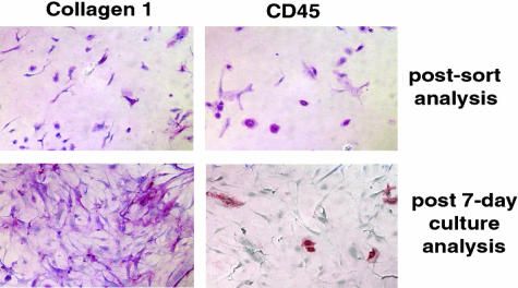
Bone marrow-derived cells are known to play important roles in repair/regeneration of injured tissues, but their roles in pathological fibrosis are less clear. Here, we report a critical role for the chemokine receptor CCR2 in the recruitment and activation of lung fibrocytes (CD45(+), CD13(+), collagen 1(+), CD34(-)). Lung fibrocytes were isolated in significantly greater numbers from airspaces of fluorescein isothiocyanate-injured CCR2(+/+) mice than from CCR2(-/-) mice. Transplant of CCR2(+/+) bone marrow into CCR2(-/-) recipients restored recruitment of lung fibrocytes and susceptibility to fibrosis. Ex vivo PKH-26-labeled CCR2(+/+) lung fibrocytes also migrated to injured airspaces of CCR2(-/-) recipients in vivo. Isolated lung fibrocytes expressed CCR2 and migrated to CCL2, and CCL2 stimulated collagen secretion by lung fibrocytes. Fibrocytes could transition into fibroblasts in vitro, and this transition was associated with loss of CCR2 expression and enhanced production of collagen 1. This is the first report describing expression of CCR2 on lung fibrocytes and demonstrating that CCR2 regulates both recruitment and activation of these cells after respiratory injury.


Figures

Figure 1
Figure 1
Cells with both mesenchymal and leukocyte markers accumulate in the airspaces of FITC-injured mice. a: C57BL/6 mice were injected with FITC on day 0 and BAL was performed on day 3 after FITC. Lavage cells were cultured for 14 days and stained for expression of col 1, CD45, or CD13. The negative control is a mixture of rabbit and rat irrelevant Ig. Positive cells were identified with alkaline phosphatase activity (red color). Staining pattern was similar for cells isolated on all other days after FITC. b: Dual-IHC staining demonstrated the presence of both mesenchymal (col 1, stained with ACMA; blue color) and leukocyte (CD45, stained with PE; red color) markers on the same cells cultured from day 5 BAL of FITC-treated mice. Data in a and b are representative of three experiments per time point in two different genetic backgrounds. c: Absolute numbers of CD45+, col 1+ cells isolated from BAL cultures on various days after FITC (n = 5 per time point, representative of three similar experiments).
Figure 2
Figure 2
Lung mince cultures contain fibrocytes. a: Lung minces from unchallenged C57BL/6 mice were cultured for 14 days and stained for expression of col 1, CD45, or CD13. The negative control was a mixture of rat and rabbit irrelevant Ig. b: Flow cytometry analysis of lung mince cultures stained for surface expression of CD45 using a directly conjugated PE Ab and intracellular col I (rabbit anti-mouse) recognized with a Cy5-conjugated donkey anti-rabbit secondary Ab. c: RT-PCR analysis of extracellular matrix gene expression between fibrocytes and fibroblasts. CD45+ fibrocytes and CD45 fibroblasts were purified from lung mince cultures via magnetic separation. Total RNA was prepared from each group of cells and RT-PCR performed for col I, col III, and fibronectin gene expression. d: Flow cytometry analysis of magnetically sorted CD45+ fibrocytes from C57BL/6 lung mince cultures pooled from three mice. After a single round of magnetic purification, fibrocytes were stained with CD45-PE (B), col 1 and donkey anti-rabbit FITC secondary (C), or with Col 1-FITC and CD34PE (D). All panels are representative of at least three separate experiments.
Figure 3
Figure 3
Lung fibrocytes express functional CCR2 receptors and migrate to CCL2 in vitro. a: Superarray analysis of chemokine receptor gene expression in fibrocytes from B6129/F2 mice. b: CCR2 IHC on cytospins of fibrocytes (CD45+) and fibroblasts (CD45) purified from n = 3 pooled lung minces. Red alkaline phosphatase staining represents positive staining. Only the CD45+ population expressed CCR2. Data are representative of three experiments. c: CD45+ fibrocytes and CD45 fibroblasts were purified from lung mince cultures and in vitro chemotaxis assays were performed. Fibronectin was used as a positive control for mesenchymal cell migration (n = 6 per condition and represents three similar experiments).
Figure 4
Figure 4
Fibrocyte chemokine receptor profiles do not change in response to FITC. Fibrocytes were purified via magnetic sorting from day 14 lung mince cultures from saline- or FITC-treated C57BL/6 mice. Receptor profiles did not change after FITC administration. Blots shown represent RNA pooled from three mice and were repeated two times in this background strain. Similarly, the profile of chemokine receptors seen on fibrocytes purified from B6129/F2 mice were not different from the profile seen in Figure 3a (not shown). RPL13A is ribosomal protein 13A.
Figure 5
Figure 5
Lung fibrocytes migrate to injured airspaces of CCR2+/+ mice in greater numbers than in CCR2−/− mice, and these differences do not reflect proliferative changes in culture. a: C57BL/6, B6/129F2, or CCR2−/− mice on either background were injected with FITC on day 0. On day 4 after FITC, BAL was performed and cell pellets were cultured and analyzed for dual expression of CD45 and col 1. Data represent n = 4 mice per group and are representative of three separate experiments. b: Fibrocytes and fibroblasts were purified from pooled lung mince cultures of day 7 FITC-treated CCR2+/+ or CCR2−/− mice. Equal numbers of cells were plated and cultured for 48 hours before the addition of 3H-thymidine for a final 16 hours. Although fibrocytes are less proliferative than fibroblasts, no differences were seen in the proliferative capacity of these cells from CCR2+/+ or CCR2−/− mice at any time point tested (24 to 72 hours, n = 6). Representative of three separate experiments.
Figure 6
Figure 6
CCR2+/+ BMT into CCR2−/− mice restores lung fibrocyte recruitment and fibrotic susceptibility. a: Wild-type B6/129F2 CCR2+/+, CCR2−/−, or CCR2−/− mice that had received a BMT with CCR2+/+ bone marrow were injected with FITC. BAL was harvested on day 4 after FITC for culture and enumeration of CD45+, col 1+ cells. The lung fibrocytes isolated from CCR2+/+ BMT mice were CCR2-positive by IHC; RT-PCR confirms that they were of the CCR2+/+ donor origin. Data represent n = 5 per group and were repeated twice. b: CCR2−/− mice were given BMT from CCR2−/− or CCR2+/+ donors and rested for 7 weeks. Mice were then injected with saline or FITC. Lung hydroxyproline content was determined 21 days later. Data represent five mice per group in the saline injections and eight mice per group in the FITC injections. All experiments were repeated two times. c: PKH-26-labeled CCR2+/+ fibrocytes were visualized in FITC-treated lungs within 24 hours of tail vein infusion.
Figure 7
Figure 7
CCL2 and TGF-β1 stimulate collagen production in lung fibrocytes. Sorted populations of lung fibrocytes (a and b) and fibroblasts (c and d) were serum-starved for 24 hours before the addition of fresh serum-free media or serum-free media + IL-13 (10 ng/ml), CCL2 (10 ng/ml), or TGF-β1 (2 ng/ml) for an additional 24 hours. Col 1 protein was analyzed by Western blot. Two blots, representative of four total blots are shown for each cell type (a and c). Blots were stripped and reprobed with β-tubulin. Densitometry was performed to normalize col I expression to β-tubulin. In each experiment, the value for serum-free media was normalized to 1 and statistical analyses were performed (b and d). CCL2 and TGF-β1 both significantly stimulated the production of col I in fibrocytes. IL-13 and TGF-β1 both stimulated the production of col I in fibroblasts.
Figure 8
Figure 8
Fibrocytes transition into fibroblasts in vitro. Fibrocytes were purified via two rounds of magnetic selection and the final purified population was greater than 99.5% positive for col I and CD45 after sorting. Purified fibrocytes were cultured for 7 days in complete media and reanalyzed for expression of col I and CD45 via IHC using alkaline phosphatase-coupled secondary reagents. After 1 week of culture, the purified fibrocytes retained col I expression, but lost CD45 expression suggesting a transition from the fibrocyte to the fibroblast phenotype. Similar results were obtained in three separate experiments.

References

    1. Thannickal VJ, Toews GB, White E, Lynch JI, Martinez F. Mechanisms of pulmonary fibrosis. Annu Rev Med. 2004;55:395–471. - PubMed
    1. Lynch JI, Toews G: Idiopathic pulmonary fibrosis. Fishman A, Pulmonary Diseases and Disorders. Edited by Fishman A. Philadelphia PA, 1997, pp 1069-1084
    1. Kuhn C. Pathology. Phan S, Thrall R, editors. New York: Marcek Dekker, Inc.,; Pulmonary Fibrosis. 1995:pp 59–83.
    1. King TE, Jr, Schwarz MI, Brown K, Tooze JA, Colby TV, Waldron JA, Jr, Flint A, Thurlbeck W, Cherniack RM. Idiopathic pulmonary fibrosis: relationship between histopathologic features and mortality. Am J Respir Crit Care Med. 2001;164:1025–1032. - PubMed
    1. Coultas D, Zumwalt R, Black W, Sobonya R. The epidemiology of interstitial lung disease. Am J Respir Crit Care Med. 1994;150:967–972. - PubMed

Publication types

MeSH terms