

An experimental model of autoimmune pancreatitis in the rat
- PMID: 15743785
- PMCID: PMC1602363
- DOI: 10.1016/S0002-9440(10)62294-8
An experimental model of autoimmune pancreatitis in the rat
Abstract
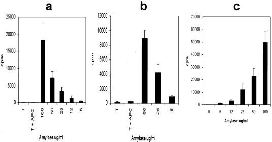
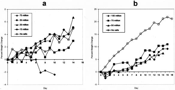
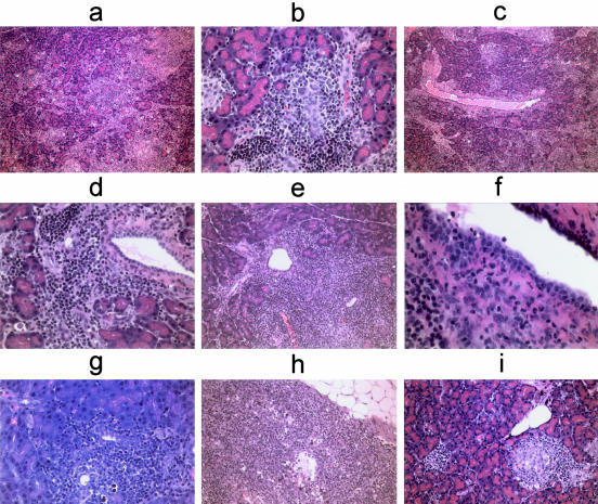
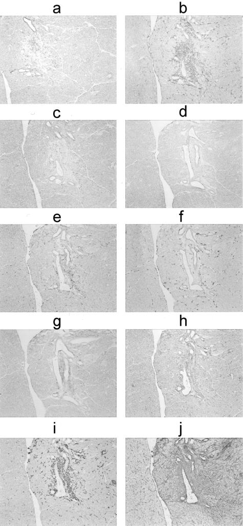
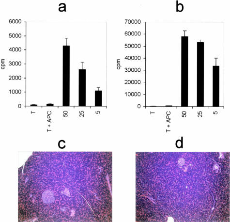
Autoimmune pancreatitis (AIP), a recently defined disease of unknown etiology, is characterized by inflammatory infiltrates in the pancreas with conspicuous involvement of the ducts. The disease clinically manifests in humans as epigastric pain, weight loss, and jaundice. This report describes the development of a novel animal model of this disease in the rat, which we have termed experimental autoimmune pancreatitis. Adoptive transfer of amylase-specific CD4(+) T cells was able to confer pancreatitis to naive syngeneic recipient animals. No treatments before the adoptive transfer of T cells were necessary for disease to ensue, and the severity of disease was proportional to the number of T cells administered. The pancreatic lesions of rats with experimental autoimmune pancreatitis were characterized histologically as overwhelmingly lymphocytic with occasional plasma cells, neutrophils, and mast cells. Acinar tissue destruction and ductular inflammation were common features, with less frequent involvement of larger ducts. Immunohistochemical analysis revealed the presence of CD4(+) T cells in large numbers as well as CD8(+) T cells, macrophages, and dendritic cells. Expression of MHC I and MHC II also increased at the site of the lesion. Clinically, the disease manifested as either failure to gain weight at a rate concomitant with control animals or as outright weight loss. Thus, administration of activated CD4(+) T cells specific for the pancreatic enzyme amylase can induce pancreatitis in the rat in a manner that is reminiscent of human AIP.
Figures
Similar articles
-
Induction of experimental autoimmune keratitis by adoptive transfer of human corneal antigen-specific T-cell line.Invest Ophthalmol Vis Sci. 2000 Dec;41(13):4182-8. Invest Ophthalmol Vis Sci. 2000. PMID: 11095613
-
Early rise in inflammation and microcirculatory disorder determine the development of autoimmune pancreatitis in the MRL/Mp-mouse.Am J Physiol Gastrointest Liver Physiol. 2008 Dec;295(6):G1274-80. doi: 10.1152/ajpgi.90341.2008. Epub 2008 Oct 30. Am J Physiol Gastrointest Liver Physiol. 2008. PMID: 18974312
-
Autoimmune pancreatitis: a systemic immune complex mediated disease.Am J Surg Pathol. 2006 Dec;30(12):1537-45. doi: 10.1097/01.pas.0000213331.09864.2c. Am J Surg Pathol. 2006. PMID: 17122509
-
Autoimmune pancreatitis: an underdiagnosed autoimmune disease with clinical, imaging and serological features.Autoimmun Rev. 2010 Feb;9(4):237-40. doi: 10.1016/j.autrev.2009.07.003. Epub 2009 Jul 18. Autoimmun Rev. 2010. PMID: 19622398 Review.
-
Histopathology of autoimmune pancreatitis: recognized features and unsolved issues.J Gastrointest Surg. 2005 Jan;9(1):6-10. doi: 10.1016/j.gassur.2004.08.010. J Gastrointest Surg. 2005. PMID: 15773046 Review. No abstract available.
Cited by
-
Deciphering autoimmune pancreatitis, a great mimicker: case report and review of the literature.Case Rep Gastrointest Med. 2015;2015:924532. doi: 10.1155/2015/924532. Epub 2015 Jan 29. Case Rep Gastrointest Med. 2015. PMID: 25705529 Free PMC article.
-
Autoimmune pancreatitis-related diabetes: quantitative analysis of endocrine islet cells and inflammatory infiltrate.Virchows Arch. 2010 Sep;457(3):329-36. doi: 10.1007/s00428-010-0948-y. Epub 2010 Jul 15. Virchows Arch. 2010. PMID: 20632032
-
The Wistar Bonn Kobori rat, a unique animal model for autoimmune pancreatitis with extrapancreatic exocrinopathy.Clin Exp Immunol. 2008 Apr;152(1):1-12. doi: 10.1111/j.1365-2249.2008.03588.x. Epub 2008 Feb 14. Clin Exp Immunol. 2008. PMID: 18279444 Free PMC article.
-
Amylase alpha-2A autoantibodies: novel marker of autoimmune pancreatitis and fulminant type 1 diabetes.Diabetes. 2009 Mar;58(3):732-7. doi: 10.2337/db08-0493. Epub 2008 Nov 10. Diabetes. 2009. PMID: 19001184 Free PMC article.
-
The Risk of Chronic Pancreatitis in Patients with Psoriasis: A Population-Based Cohort Study.PLoS One. 2016 Jul 28;11(7):e0160041. doi: 10.1371/journal.pone.0160041. eCollection 2016. PLoS One. 2016. PMID: 27467265 Free PMC article.
References
-
- Pearson RK, Longnecker DS, Chari ST, Smyrk TC, Okazaki K, Frulloni L, Cavallini G. Controversies in clinical pancreatology: autoimmune pancreatitis: does it exist? Pancreas. 2003;27:1–13. - PubMed
-
- Kloppel G, Luttges J, Lohr M, Zamboni G, Longnecker D. Autoimmune pancreatitis: pathological, clinical, and immunological features. Pancreas. 2003;27:14–19. - PubMed
-
- Kamisawa T, Egawa N, Nakajima H. Autoimmune pancreatitis is a systemic autoimmune disease. Am J Gastroenterol. 2003;98:2811–2812. - PubMed
-
- Kamisawa T, Funata N, Hayashi Y, Eishi Y, Koike M, Tsuruta K, Okamoto A, Egawa N, Nakajima H. A new clinicopathological entity of IgG4-related autoimmune disease. J Gastroenterol. 2003;38:982–984. - PubMed
-
- Kamisawa T, Egawa N, Nakajima H, Tsuruta K, Okamoto A, Kamata N. Clinical difficulties in the differentiation of autoimmune pancreatitis and pancreatic carcinoma. Am J Gastroenterol. 2003;98:2694–2699. - PubMed
MeSH terms
Substances
LinkOut - more resources
Full Text Sources
Other Literature Sources
Medical
Research Materials
Miscellaneous
