. 2005 Mar;166(3):793-800.
doi: 10.1016/S0002-9440(10)62300-0.

Vascular adhesion protein-1 is involved in both acute and chronic inflammation in the mouse

Affiliations

Vascular adhesion protein-1 is involved in both acute and chronic inflammation in the mouse

Marika Merinen et al. Am J Pathol. 2005 Mar.

Abstract

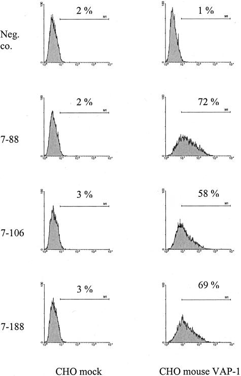
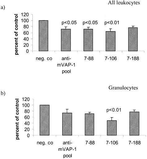
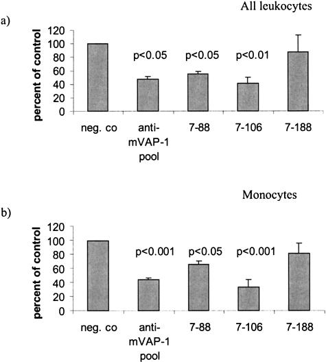
Vascular adhesion protein-1 (VAP-1) is an endothelial molecule that possesses both adhesive and enzymatic properties in vitro. So far, however, elucidation of its in vivo function has suffered from the lack of function-blocking reagents that are suitable for use in animal models. In this work we produced monoclonal antibodies against murine VAP-1 and characterized them using in vitro binding assays. We then examined whether the antibodies could prevent leukocyte migration in in vivo inflammation models, including two acute models (peritonitis induced with proteose peptone and interleukin-1 and air pouch inflammation enhanced by CCL21) and one chronic model (autoimmune diabetes in nonobese diabetic mice). Antibodies 7-88 and 7-106 inhibited migration of granulocytes and monocytes in both acute models of inflammation. Strikingly, antibody 7-88 significantly prevented diabetes in a subset of nonobese diabetic mice. The results show for the first time that in mouse models of inflammation, VAP-1 mediates leukocyte trafficking to sites of inflammation and thus is a potential target for anti-inflammatory therapies.


Figures

Figure 1
Figure 1
Reactivity of anti-VAP-1 antibodies against recombinant VAP-1. Antibodies (7-88, 7-106, 7-188, and a negative control antibody) were used to stain both CHO cells transfected with a cDNA encoding mouse VAP-1 or with vector only (CHO mock). The x axis is the fluorescence intensity in a logarithmic scale and the y axis is relative number of cells. The experiment has been repeated four times.
Figure 2
Figure 2
VAP-1 is present in a subpopulation of vessels in several organs and in smooth muscle cells. Frozen sections of the indicated organs were stained with 7-88 anti-VAP-1 antibody or a negative control antibody as the first stage reagent followed by peroxidase-conjugated anti-rat Ig. Arrows point out some positive vessels. Sm, smooth muscle. Original magnifications, ×200.
Figure 3
Figure 3
VAP-1-dependent inhibition of lymphocytes to lymph node HEVs and to vasculature in inflamed pancreas. Lymphocyte binding to lymph node HEV (a) and vessels in inflamed pancreata (b) was measured using a Stamper-Woodruff-type of an adhesion assay. The results are presented as percentage of control binding (set as 100% in the presence of a negative control antibody) ±SEM. c: An example of lymphocyte binding to two HEV-like vessels (outlined by dashed lines) in an inflamed pancreatic islet (the border of the islet is outlined with a dotted line). Arrows point out some of the bound lymphocytes (dark round cells). Original magnifications, ×200.
Figure 4
Figure 4
VAP-1 contributes to granulocyte trafficking in inflamed peritoneum. Proteose peptone and interleukin-1α were used to induce peritonitis and at 1 hour and 4 hours thereafter anti-VAP-1 or control mAbs were administered intravenously. The lavage fluid was collected at 18 hours after induction and the infiltrated leukocytes and granulocytes were counted. The results are presented as percentages of the number of total leukocytes (a) and granulocytes (b) recovered from the mice treated with the control antibody (set as 100%) ±SEM.
Figure 5
Figure 5
VAP-1 inhibits leukocyte trafficking to inflamed air pouch. The mice were treated after onset of the inflammation with the indicated antibodies. At the end of the experiment the cells in the lavage fluid were collected and counted. The results are presented as percentages of the number of leukocytes (a) and monocytes (b) recovered from the mice treated with the control antibody (set as 100%) ±SEM.
Figure 6
Figure 6
VAP-1 is present on the surface of pancreatic vessel wall at an early age in NOD mice. The mice were intravenously injected with anti-VAP-1 antibody (7-88) or a negative control antibody. The bound antibody was detected in frozen sections using a FITC-conjugated second stage antibody. Original magnifications, ×400.
Figure 7
Figure 7
VAP-1 is a target for the prevention of autoimmune diabetes. NOD mice were treated intraperitoneally for 6 months twice per week with anti-VAP-1 antibody (7-88) or an irrelevant class-matched control antibody and the incidence of diabetes was followed for 1 year. See Materials and Methods for further details.
Figure 8
Figure 8
L-selectin phenotypes after antibody treatments. Starting at the age of 3 weeks, NOD mice were treated for 10 weeks with 7-88 or a negative control antibody twice per week. At the end of the experiment the lymphocytes were isolated from the indicated organs and double stained. Each histogram is a pooled cell suspension of either anti-VAP-1-treated animals (eight mice) or control-treated ones (eight mice). The x axis is the fluorescence intensity in a logarithmic scale and the y axis is the relative number of cells. The black line is anti-VAP-1-treated animals and the gray line illustrates control-treated mice.

Similar articles

Cited by

References

    1. Salmi M, Yegutkin G, Lehvonen R, Koskinen K, Salminen T, Jalkanen S. A cell surface amine oxidase directly controls lymphocyte migration. Immunity. 2001;14:265–276. - PubMed
    1. Butcher EC. Leukocyte-endothelial cell recognition: three (or more) steps to specificity and diversity. Cell. 1991;67:1033–1036. - PubMed
    1. Salmi M, Jalkanen S. VAP-1: an adhesin and an enzyme. Trends Immunol. 2001;22:211–216. - PubMed
    1. Jaakkola K, Nikula T, Holopainen R, Vähäsilta T, Matikainen M-T, Laukkanen M-L, Huupponen R, Halkola L, Nieminen L, Hiltunen J, Parviainen S, Clark MR, Knuuti J, Savunen T, Kääpä P, Voipio-Pulkki L-M, Jalkanen S. In vivo detection of vascular adhesion protein-1 in experimental inflammation. Am J Pathol. 2000;157:463–471. - PMC - PubMed
    1. Tohka S, Laukkanen M, Jalkanen S, Salmi M. Vascular adhesion protein 1 (VAP-1) functions as a molecular brake during granulocyte rolling and mediates recruitment in vivo. FASEB J. 2001;15:373–382. - PubMed

Publication types

MeSH terms