. 2005 Mar;166(3):811-22.
doi: 10.1016/S0002-9440(10)62302-4.

Abnormal T-cell reactivity against paternal antigens in spontaneous abortion: adoptive transfer of pregnancy-induced CD4+CD25+ T regulatory cells prevents fetal rejection in a murine abortion model

Affiliations

Abnormal T-cell reactivity against paternal antigens in spontaneous abortion: adoptive transfer of pregnancy-induced CD4+CD25+ T regulatory cells prevents fetal rejection in a murine abortion model

Ana Claudia Zenclussen et al. Am J Pathol. 2005 Mar.

Abstract

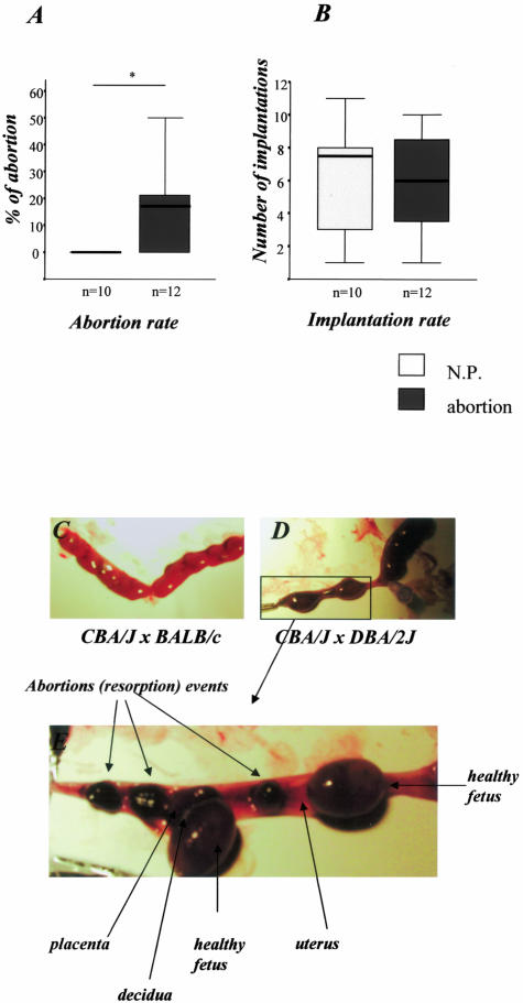
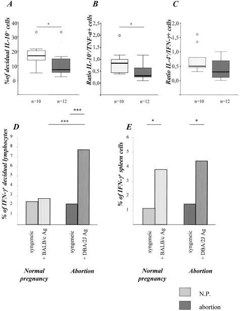
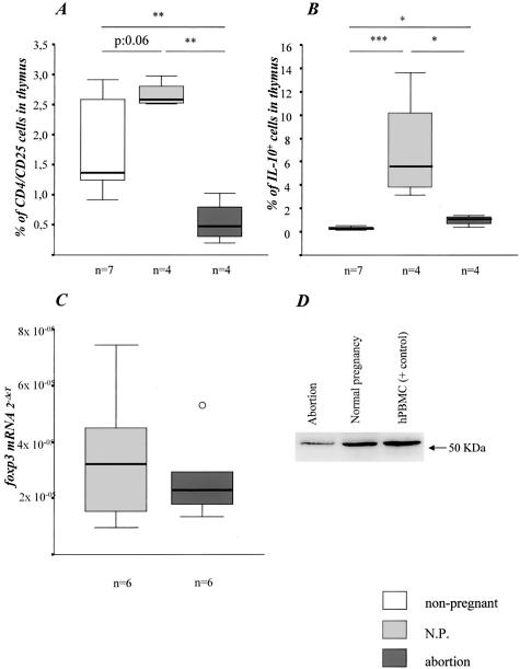
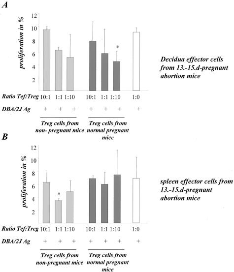
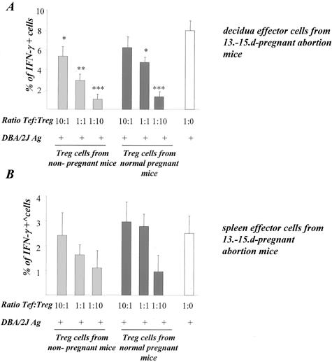
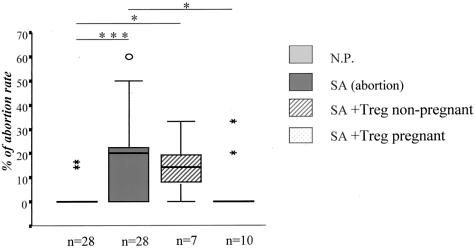
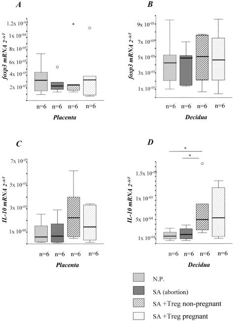
Mammalian pregnancy is thought to be a state of immunological tolerance. The mechanisms underlying this phenomenon are still poorly understood. Here, we determined whether an inappropriate function of T regulatory (Treg) cells is involved in the pathogenesis of spontaneous abortion. We evaluated spleen and decidual lymphocytes from CBA/J mice undergoing immunological abortion (DBA/2J-mated) or having normal pregnancy (BALB/c-mated) on day 14 of gestation for ex vivo cytokine production after PMA or paternal antigen (alloantigen) stimulation. Treg activity was characterized by quantifying CD4(+)CD25(+) cells, foxp3 expression, and interleukin-10 secretion. Decidual lymphocytes from abortion CBA/J mice contained a significantly higher frequency of interferon-gamma-producing T cells specific for paternal antigens compared to those from normal pregnancy (7.8% versus 2.7%, P < 0.05). Compared to virgin CBA/J females, normal pregnant mice showed strongly elevated numbers of CD4(+)CD25(+) and interleukin-10(+) Treg cells in the thymus whereas significantly lower frequencies of Treg cells were observed in abortion mice. Very interestingly, CD4(+)CD25(+) Treg cells from normal pregnant and nonpregnant CBA/J mice could inhibit both proliferation and interferon-gamma secretion of lymphocytes from abortion mice in vitro whereas in vivo prevention of fetal rejection could only be achieved after adoptive transfer of Treg cells from normal pregnant mice. Our data suggest that pregnancy-induced Treg cells play a vital role in maternal tolerance to the allogeneic fetus.


Figures

Figure 1
Figure 1
Abortion rates. A: DBA/2J-mated CBA/J mice (abortion mice) showed enhanced abortion rates compared to control mice (CBA/J × BALB/c, normal pregnant, N.P.) as analyzed by the nonparametric Mann-Whitney U-test (P < 0.05) in two independent experiments. B: Comparable implantation rates between both experimental groups. The data are presented as median ± 75% quartiles. C and D: Representative pictures of uteri from normal pregnant mice (C) and from abortion mice (D). E: The left uterus from abortion mice is amplified and five implantation sites can be observed. Three of them are abortions whereas the other two are normal implantation sites where healthy fetuses and placentas can be perfectly identified from each other.
Figure 2
Figure 2
Cytokine production. Abortion mice produced significantly less IL-10 (A) and presented therefore a statistically significant diminution in the IL-10/TNF-α ratio when compared to normal pregnant mice (B, P < 0.05) as analyzed by the nonparametric Mann-Whitney U-test (P < 0.05). The IL-4/IFN-γ (C) ratio was slightly but not significantly diminished as analyzed in decidual cells after ex vivo stimulation with PMA/ionomycin. The data are presented as median ± 75% quartiles. D and E: Ag-specific cytokine production. Decidual (D) but not spleen (E) cells from abortion mice secreted more IFN-γ when stimulated with male APCs compared to normal pregnant mice (P < 0.001). IFN-γ production was analyzed in pooled decidual (D) or spleen (E) immune cells from at least three normal pregnant or abortion mice when co-cultured with CBA/J antigen (syngeneic control) or male antigens (BALB/c or DBA/2J). The data are representative of two independent experiments. Statistical significances were analyzed by the χ2 test and α-adjusted by Bonferroni’s test.
Figure 3
Figure 3
Treg activity. A: Normal pregnant mice presented more Treg cells than nonpregnant mice (P = 0.06). DBA/2J-mated CBA/J females undergoing abortion showed significantly decreased numbers of Treg cells when compared to BALB/c-mated CBA/J females (P < 0.01). B: Thymocytes from normal pregnant mice produced significantly more IL-10 when compared to nonpregnant (P < 0.001) or normal pregnant mice (P < 0.05). The data are presented as median ± 75% quartiles. Significances were calculated using the nonparametric Kruskall-Wallis test followed by the Mann-Whitney U-test. Abortion mice presented lower placental foxp3 mRNA (C) and protein levels (D) as analyzed by real-time RT-PCR and Western blot, respectively. In Western blot human PBMCs were used as a positive control.
Figure 4
Figure 4
Treg modulated the in vitro proliferation. A to C: The results of our in vitro assays by co-culturing pooled decidual (A) or spleen (B) effector cells with Treg cells isolated from nonpregnant (gray bars) or normal pregnant mice (black bars) and male antigens (DBA/2J APCs). White bars show the proliferation controls without Treg cells addition. Pooled effector cells were stained with CFSE and their proliferation was analyzed 3 days after culture. The addition of Treg cells led to a significant inhibition in the proliferation rates by decidual cells (A) and a slight diminution by spleen cells (B). Data are a mean of three independent experiments, in which pooled cells from at least three animals were analyzed. Statistical significances were analyzed by the χ2 test and α-adjusted by Bonferroni’s test.
Figure 5
Figure 5
Treg modulated the in vitro IFN-γ production. The addition of Treg cells led to a significant inhibition in the IFN-γ production by effector cells. The in vitro assays were performed by co-culturing pooled decidual (A) or spleen (B) effector cells with Treg cells isolated from nonpregnant (gray bars) or normal pregnant mice (black bars) and male antigens (DBA/2J APCs). White bars show IFN-γ secretion controls without Treg cell addition. IFN-γ secretion was analyzed 3 days after culture. Data are a mean of three independent experiments, in which pooled cells from at least three animals were analyzed. Statistical significances were analyzed by the χ2 test and α-adjusted by Bonferroni’s test.
Figure 6
Figure 6
NP-Treg transfer avoided abortion. The injection of 2 × 10⁵ Treg cells from normal pregnant, but not from nonpregnant mice led to a statistically significant diminution in the abortion rate (P < 0.05). The abortion rate from NP-Treg cell-transferred mice was comparable to the normal pregnant controls. The data are presented as median ± 75% quartiles and the statistically significant differences were analyzed by Kruskall-Wallis test followed by Mann-Whitney U-test.
Figure 7
Figure 7
Treg transfer up-regulated foxp3 and IL-10 mRNA. Abortion mice presented diminished or comparable foxp3 (A and B) and IL-10 (C and D) mRNA levels in placenta and decidua when compared to normal pregnant mice. The injection of 2 × 10⁵ Treg cells from both normal pregnant or nonpregnant mice led to augmented foxp3 (slightly) and IL-10 mRNA levels (significantly). The data are presented as median ± 75% quartiles and the statistically significant differences were analyzed by Kruskall-Wallis test followed by Mann-Whitney U-test.
Figure 8
Figure 8
Treg transfer up-regulated the expression of progesterone receptors and had no effect on the estrogen receptor expression. A: Abortion mice presented lower PR levels than normal pregnant mice. Treg transfer restored the receptor expression to levels comparable with those observed in normal pregnant mice. B: No differences were observed between any groups regarding the expression of ER. C to F: Representative PR staining in normal pregnant mice (C), abortion-prone mice (D), and in mice transferred with Treg from nonpregnant (E) or pregnant animals (F). G is a typical field for ER staining while H depicts our negative control.

Similar articles

Cited by

References

    1. Medawar PB. Some immunological and endocrinological problems raised by the evolution of viviparity in vertebrates. Symp Soc Exp Biol. 1953;44:320–338.
    1. Tafuri A, Alferink J, Moller P, Hammerling G, Arnold B. T cell awareness of paternal alloantigens during pregnancy. Science. 1995;270:630–633. - PubMed
    1. Munn D, Zhou M, Attwood J, Bondarev I, Conway S, Marshall B, Brown C, Mellor A. Prevention of allogeneic fetal rejection by tryptophan catabolism. Science. 1998;281:1191–1193. - PubMed
    1. Rouas-Freiss N, Goncalves RM, Menier C, Dausset J, Carosella ED. Direct evidence to support the role of HLA-G in protecting the fetus from maternal uterine natural killer cytolysis. Proc Natl Acad Sci USA. 1997;94:11520–11525. - PMC - PubMed
    1. Makriagiannis A, Zoumakis E, Kalantaridou C, Coutifaris C, Margioris AN, Coukos G, Rice KC, Gravansi A, Chrousos GP. Corticotropin-releasing hormone promotes blastocytes implantation and early maternal tolerance. Nat Immunol. 2001;18:367–391. - PubMed

Publication types

MeSH terms