. 2005 Mar;166(3):843-55.
doi: 10.1016/S0002-9440(10)62305-X.

A cyclosporine-sensitive psoriasis-like disease produced in Tie2 transgenic mice

Affiliations

A cyclosporine-sensitive psoriasis-like disease produced in Tie2 transgenic mice

Daniel Voskas et al. Am J Pathol. 2005 Mar.

Abstract

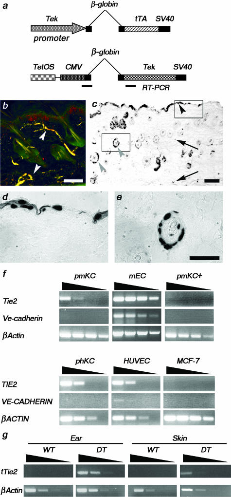
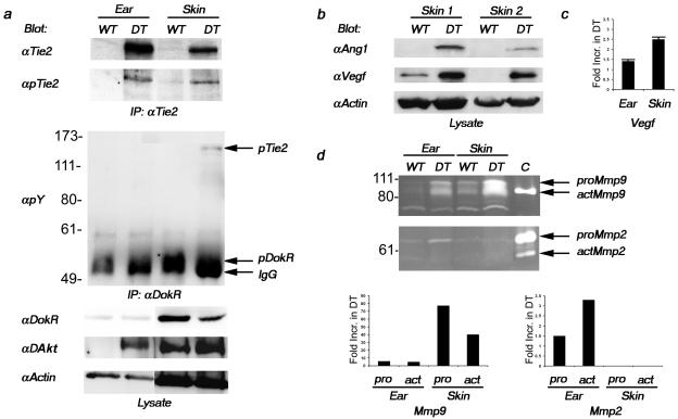
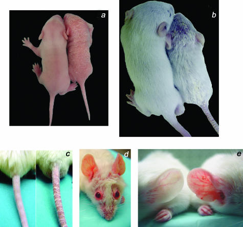
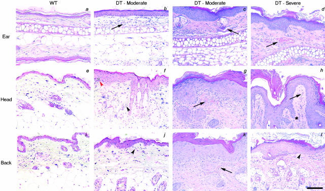
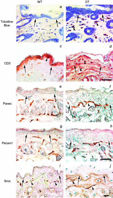
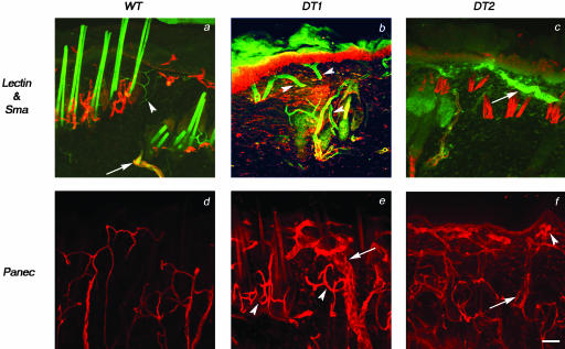
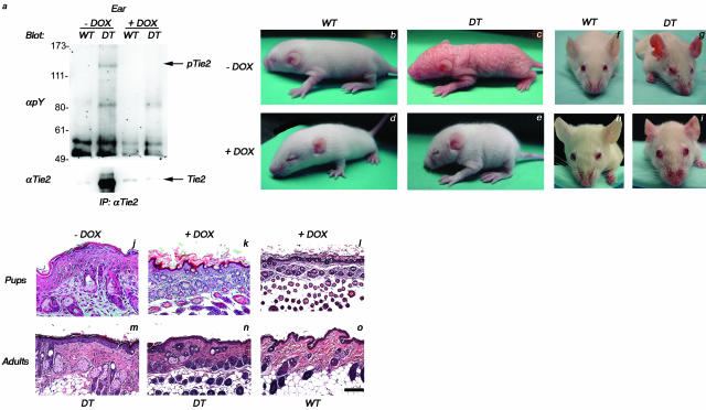
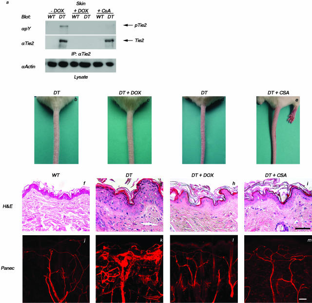
Psoriasis is a common, persistent skin disorder characterized by recurrent erythematous lesions thought to arise as a result of inflammatory cell infiltration and activation of keratinocyte proliferation. Unscheduled angiogenic growth has also been proposed to mediate the pathogenesis of psoriasis although the cellular and molecular basis for this response remains unclear. Recently, a role for the angiopoietin signaling system in psoriasis has been suggested by studies that demonstrate an up-regulation of the tyrosine kinase receptor Tie2 (also known as Tek) as well as angiopoietin-1 and angiopoietin-2 in human psoriatic lesions. To examine temporal expression of Tie2, we have developed a binary transgenic approach whereby expression of Tie2 can be conditionally regulated by the presence of tetracycline analogs in double-transgenic mice. A psoriasis-like phenotype developed in double-transgenic animals within 5 days of birth and persisted throughout adulthood. The skin of affected mice exhibited many cardinal features of human psoriasis including epidermal hyperplasia, inflammatory cell accumulation, and altered dermal angiogenesis. These skin abnormalities resolved completely with tetracycline-mediated suppression of transgene expression, thereby illustrating a complete dependence on Tie2 signaling for disease maintenance and progression. Furthermore, the skin lesions in double-transgenic mice markedly improved after administration of the immunosuppressive anti-psoriatic agent cyclosporine, thus demonstrating the clinical significance of this new model.


Figures

Figure 1
Figure 1
Tie2 expression in DT mice. a: Schema of the two transgenes used in these studies to produce the driver line (top) and the responder line (bottom). b: Panec (green) and Tie2 (red) co-staining on a section from adult skin demonstrates co-localization (yellow) of Tie2 with an endothelial cell marker (white arrowheads). c–e: Analysis of Tie2-driven LacZ expression in the skin of DT mice. Expression of the nlsLacZ transgene is seen in endothelial cells (arrows), keratinocytes (black arrowhead), and epithelial cells of hair follicles (gray arrowhead) (c). Increased magnification of insets in c show keratinocytes (d) and epithelial cells of hair follicles (e). f: Semiquantitative RT-PCR analysis of Tie2 and Ve-cadherin expression was completed on RNA isolated from primary mouse (pm) and human (ph) KCs. Tie2 expression is down-regulated in pmKCs on treatment with calcium (pmKC+). Mouse endothelioma cells and human umbilical vein endothelial cells are positive whereas MCF-7 cells are negative for Tie2 and Ve-cadherin expression. The βActin control demonstrates the presence of amplifiable RNA in all samples. cDNA templates were undiluted, diluted 1 in 10, 1 in 100, or 1 in 1000, as illustrated by the wedge. g: Semiquantitative RT-PCR analysis of the Tie2 transgene was performed on RNA isolated from the ear and skin of DT and normal or WT adult mice. Expression of the Tie2 transgene can be readily detected in the ear and skin of a DT adult mouse. The βActin control indicates the presence of amplifiable RNA. cDNA templates were diluted as above. Scale bars, 100 μm.
Figure 2
Figure 2
Increased Tie2 expression and signal transduction in DT mice. a: Tie2 immunoprecipitates from lysates extracted from the ear and skin of DT and normal (WT) adult mice were immunoblotted with antibodies recognizing phosphorylated Tie2 (pTie2). The blot was then reprobed with anti-Tie2 antibodies. Increased expression and phosphorylation of Tie2 can be observed in DT mice. Lysates were also immunoprecipitated with anti-DokR antibodies and immunoblotted with antibodies recognizing phosphotyrosine (pY). The blot was then reprobed with antibodies recognizing DokR. IgG indicates the position of IgG. Nonimmunoprecipitated lysates (lysate) were incubated with antibodies recognizing either phosphorylated Akt (pAkt) or Actin. b: Nonimmunoprecipitated lysates extracted from the skin of two different pairs of DT and WT adult mice were immunoblotted with antibodies recognizing Ang1, Vegf, or Actin. c: Vegf protein levels as determined by enzyme-linked immunosorbent assay from lysates prepared from the ear and skin of three different pairs of animals. The fold-increase in Vegf expression in DT over normal mice was determined by densitometry. d: Gelatin zymography was performed on lysates prepared from the ear and skin of DT and WT mice. The positions of the 92-kd pro- (proMmp9) and 84- and 82-kd active (actMmp9) forms of Mmp9 are indicated as well as the 72-kd pro- (proMmp2) and 62-kd active (actMmp2) forms of Mmp2. C indicates the position of the positive controls. The fold-increase in Mmp activity in DT over normal mice was determined by densitometry of this representative experiment.
Figure 3
Figure 3
Excess Tie2 expression induces skin abnormalities in DT mice. a: A DT mouse (right) and a normal (WT) littermate (left) at 7 days of age. Erythema and scaling can be observed on the head, back, and tail of the affected mouse. b: A DT mouse (right) and a normal littermate (left) at 2 weeks of age. Note the abnormal appearance of the fur with superficial scaling on the head, lower back, and tail. c: Prominent scaling and redness on the tail of a DT mouse (right) compared to a normal littermate (left) at 2 weeks of age. d: A DT adult mouse with prominent redness around the eyes, ears, and snout. e: Comparison of ear skin shows increased redness and keratinization on the skin surface of DT mice that is not observed in WT littermates.
Figure 4
Figure 4
Histological analysis of the skin of DT mice. a–l: H&E staining of skin sections from the ear, head, or back of either adult WT mice (a, e, and i), or moderate (b, c, f, g, j, and k) or severely affected (d, h, and l) DT mice. Note the progression in epidermal thickening from moderately affected skin to severely affected skin and the extension of structures resembling rete pegs in the dermis in some regions (asterisk in h). Also note the increased vessel area density (arrows) in DT animals in the dermal papillae with lymphocytic infiltration (black arrowheads). Pockets of neutrophils can also be seen in the cornified layer forming a microabscess (red arrowhead) and in other areas throughout the dermis. Scale bar, 50 μm.
Figure 5
Figure 5
Immunohistological analysis of the skin of DT mice. a and b: Toluidine blue staining of thin sections reveals infiltration of mast cells (purple staining) throughout the dermis of DT (b, arrowheads) but not WT mice (a). c and d: Staining of thin sections for the presence of CD3-positive lymphocytes illustrates an apparent increase in T cells in the subepidermal layer of DT mice (d, arrowheads) compared to WT littermates (c). e–j: Immunohistochemical staining for Panec, Pecam1, and Sma in skin sections from a DT and WT adult mouse. Staining with the endothelial cell markers Panec (e and f) and Pecam1 (g and h) reveals large vessels closer to the epidermis in DT (f and h, arrowheads) compared to WT (e and h) mice. Sma positivity marks large supported vessels in the deep dermis of both DT and WT skin (arrowheads in i and j). In all sections, the dermo-epidermal junction is indicated with arrows. Scale bars: 50 μm (a–j); 100 μm (k and l).
Figure 6
Figure 6
Increased vessel size and tortuousity in DT mice. Thick sections (100 μm) of skin from animals perfused with a fluorescein isothiocyanate-labeled lectin (green) were stained with anti-Sma or anti-Panec antibodies (red) and visualized by confocal microscopy. a: Examination of skin from WT mice reveals vertical vessels connecting the cutaneous plexus to the subpapillary plexus that gradually decrease in diameter as they extend toward the epidermis and become progressively less positive for Sma (arrow) despite remaining lectin-positive (arrowhead). Capillaries in the papillary layer of the dermis of WT mice also stain positive for lectin alone (arrowhead). b and c: Examination of skin from two DT mice reveals large vertical vessels connecting the cutaneous plexus to the subpapillary plexus that do not decrease in diameter toward the epidermis (arrowheads in b). The indicated vessel also appears to retain increased diameter on bifurcation. The small Sma-negative capillaries below the epidermis of WT skin are replaced by large vessels that remain Sma-negative in DT skin (arrow in c). d–f: The lack of Sma staining within the subpapillary plexus is not because of problems with antibody penetration because similar thick sections stained with Panec demonstrate an intricate vascular bed beneath the epidermis in both WT (d) and DT (e and f) mice. The dermal vasculature of DT skin appears more complex than that of WT skin because vessels stained with Panec are generally of greater diameter (arrow in e) and appear more tortuous, especially around hair follicles (arrowheads in e). In addition, vessels with large lumens (arrow in f) and tangled vessels (arrowhead in f) can be seen throughout the dermis. Scale bar, 50 μm.
Figure 7
Figure 7
Repression of Tie2 transgene expression with doxycycline reverses the abnormal skin phenotype seen in DT mice. a: Western blot analysis of Tie2 isolated from ears of adult mice demonstrates the reduction in Tie2 expression levels after 7 days of doxycycline (Dox) administration. b and c: A DT pup (c) displaying the abnormal skin phenotype and a WT littermate (b) at 7 days of age before doxycycline treatment. d and e: The DT (e) and WT (d) pups from above shown at 11 days of age after 4 days of doxycycline treatment. f and g: A DT adult (g) displaying the abnormal skin phenotype and an age-matched WT adult (f) before treatment with doxycycline. h and i: The DT (i) and WT (h) adults from above shown after treatment with doxycycline for 7 days. j–o: H&E staining of skin sections taken from either littermate or age-matched counterparts of the DT and WT pups (j–l) and adults (m–o) from above demonstrates that the abnormal skin seen in untreated DT pups and adults (j and m) becomes architecturally comparable to that seen in WT counterparts (l and o) on the addition of doxycycline (k and n). Note in particular the thinning of the superficial epidermis of the squamous epithelium in treated DT mice (k and n). Scale bar, 40 μm.
Figure 8
Figure 8
Resolution of the psoriasiform skin phenotype with CsA treatment. DT or WT animals were treated for 12 days with either doxycycline (DT+Dox) or CsA (DT+CsA) or with saline alone (DT). a: Western blot analysis of Tie2 isolated from skin of adult mice demonstrates the reduction in Tie2 phosphorylation levels after 12 days of doxycycline or cyclosporine administration. b–e: The scaly red appearance of the tail in untreated DT mice (b and d) is resolved in DT mice treated with either doxycycline (c) or cyclosporine (e) and the tail more closely resembles that of a WT littermate (not shown). f–i: Histological analysis (H&E) of thin sections taken from these animals illustrates pronounced epidermal thinning and reduced dermal cellularity in mice treated with either doxycycline (h) or cyclosporine (i) when compared to untreated DT mice (g) and the skin more closely resembles that of the WT counterpart (f). j–m: Analysis of blood vessel architecture as visualized by Panec staining of thick sections taken from lectin-perfused WT animals (j) or DT animals treated with either doxycycline (l) or cyclosporine (m) demonstrates a dramatic decrease in vascular density when compared to untreated DT mice (k). Scale bar, 50 μm.

Similar articles

Cited by

References

    1. Dumont DJ, Gradwohl G, Fong GH, Puri MC, Gertsenstein M, Auerbach A, Breitman ML. Dominant-negative and targeted null mutations in the endothelial receptor tyrosine kinase, tek, reveal a critical role in vasculogenesis of the embryo. Genes Dev. 1994;8:1897–1909. - PubMed
    1. Sato TN, Tozawa Y, Deutsch U, Wolburg-Buchholz K, Fujiwara Y, Gendron-Maguire M, Gridley T, Wolburg H, Risau W, Qin Y. Distinct roles of the receptor tyrosine kinases Tie-1 and Tie-2 in blood vessel formation. Nature. 1995;376:70–74. - PubMed
    1. Suri C, Jones PF, Patan S, Bartunkova S, Maisonpierre PC, Davis S, Sato TN, Yancopoulos GD. Requisite role of angiopoietin-1, a ligand for the TIE2 receptor, during embryonic angiogenesis. Cell. 1996;87:1171–1180. - PubMed
    1. Maisonpierre PC, Suri C, Jones PF, Bartunkova S, Wiegand SJ, Radziejewski C, Compton D, McClain J, Aldrich TH, Papadopoulos N, Daly TJ, Davis S, Sato TN, Yancopoulos GD. Angiopoietin-2, a natural antagonist for Tie2 that disrupts in vivo angiogenesis. Science. 1997;277:55–60. - PubMed
    1. Ward N, Dumont DJ. The angiopoietins and Tie2/Tek: adding to the complexity of cardiovascular development. Semin Cell Dev Biol. 2002;1:19–27. - PubMed

Publication types

MeSH terms