. 2005 Mar;166(3):913-21.
doi: 10.1016/S0002-9440(10)62311-5.

Up-regulation of the lymphatic marker podoplanin, a mucin-type transmembrane glycoprotein, in human squamous cell carcinomas and germ cell tumors

Affiliations

Up-regulation of the lymphatic marker podoplanin, a mucin-type transmembrane glycoprotein, in human squamous cell carcinomas and germ cell tumors

Vivien Schacht et al. Am J Pathol. 2005 Mar.

Abstract

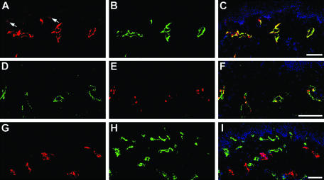
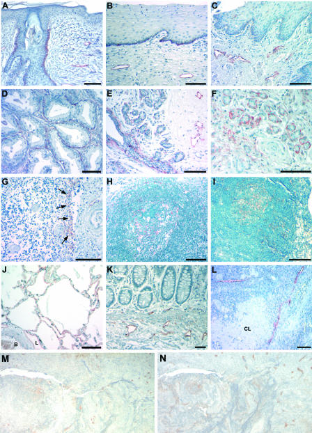
The mucin-type glycoprotein podoplanin is specifically expressed by lymphatic but not blood vascular endothelial cells in culture and in tumor-associated lymphangiogenesis, and podoplanin deficiency results in congenital lymphedema and impaired lymphatic vascular patterning. However, research into the biological importance of podoplanin has been hampered by the lack of a generally available antibody against the human protein, and its expression in normal tissues and in human malignancies has remained unclear. We generated a human podoplanin-Fc fusion protein and found that the commercially available mouse monoclonal antibody D2-40 specifically recognized human podoplanin, as assessed by enzyme-linked immunosorbent assay and Western blot analyses. We found that, in addition to lymphatic endothelium, podoplanin was also expressed by peritoneal mesothelial cells, osteocytes, glandular myoepithelial cells, ependymal cells, and by stromal reticular cells and follicular dendritic cells of lymphoid organs. These findings were confirmed in normal mouse tissues with anti-podoplanin antibody 8.1.1. Podoplanin was also strongly expressed by granulosa cells in normal ovarian follicles, and by ovarian dysgerminomas and granulosa cell tumors. Although podoplanin was primarily absent from normal human epidermis, its expression was strongly induced in 22 of 28 squamous cell carcinomas studied. These findings suggest a potential role of podoplanin in tumor progression, and they also identify the first commercially available antibody for the specific staining of a defined lymphatic marker in archival human tissue sections, thereby enabling more widespread studies of tumor lymphangiogenesis in human cancers.


Figures

Figure 1
Figure 1
The D2-40 antibody specifically recognizes human podoplanin. A and B: Rat myoblasts transiently transfected with human podoplanin-cDNA demonstrated labeling with D2-40 antibody (B, green; blue = nuclear Hoechst stain) whereas control-transfected myoblasts are negative (A). C: D2-40 antibody specifically recognizes the human podoplanin-Fc fusion protein, but not a human LYVE-1-Fc fusion protein, as detected by Western blotting. D: ELISA shows specific binding of the D2-40 antibody to immobilized human podoplanin-Fc fusion protein, as compared to low nonspecific binding to human LYVE-1-Fc fusion protein. E: Four days after anti-human podoplanin-siRNA transfection of human primary LECs, Western blots using the D2-40 antibody reveal reduced levels of endogenous podoplanin protein, using two different siRNA oligonucleotides (R1 and R2), as compared with control cells (C1, control vector cDNA; C2, sham-transfected cells). Scale bar, 50 μm (B).
Figure 2
Figure 2
Specific detection of podoplanin expression by LECs in normal human skin using the D2-40 antibody. A to C: Double-immunofluorescence stains for podoplanin (A) and for the lymphatic-specific hyaluronan receptor LYVE-1 (B) revealed co-localization on LECs (C merge, nuclei are labeled blue). In addition, podoplanin was focally expressed by basal epidermal keratinocytes (arrows). Lymphatic endothelial cells stained by D2-40 (D) also expressed the lymphatic-specific homeobox protein Prox1 (E, F merge). In contrast, double-immunofluorescence stains for podoplanin (G) and for the blood vessel-specific marker CD34 (H) demonstrated mutually exclusive expression by lymphatics and blood vessels. Scale bars, 100 μm.
Figure 3
Figure 3
Podoplanin expression in normal human tissues. In the skin (A), cervix (B), and esophagus (C), basal keratinocytes and LECs express podoplanin, whereas blood vascular endothelial cells are not labeled. Podoplanin is also expressed by fibromyocytes in the prostate (D) and by myoepithelial cells of breast glands (E) and salivary glands (F). G: Podoplanin expression by stromal cells in the medulla of the thymus near invaginations of the capsule that penetrates through the thymic cortex (arrows). H and I: Expression of podoplanin by follicular dendritic cells of germinal centers of the tonsils (H) and of lymph nodes (I). J: In the lung, alveolar type I cells and LECs (L, lymphatic vessel; B, blood vessel) express podoplanin. Lymphatic endothelial expression in the colon (K) and the ovary (L); a corpus albicans (CL) is unlabeled. M: Follicular dendritic cells express CD21 and also express podoplanin (N). Scale bars, 100 μm.
Figure 4
Figure 4
Podoplanin expression in normal mouse tissues. Immunohistochemical staining demonstrated podoplanin expression by lymphatic vessels of the skin (A) and the tongue (B). D: In the central nervous system, the ependyma lining the ventricular system and the choroid plexus expressed podoplanin. E: The perineurium demonstrated podoplanin expression around spinal nerve roots. Myoepithelial cells of the salivary gland (C) and fibromyocytes of the testis (F) also express podoplanin. In lymphoid organs, podoplanin is expressed by endothelial cells lining lymphatic vessels (L) and by follicular dendritic cells of the germinal centers in the spleen (G, arrows). H: In lymph nodes, reticulum cells of the cortex around follicles express podoplanin. Expression of podoplanin in osteocytes (I; arrows), the periosteum (P), meninges (M) and lung alveolar type I cells (J). K: Only lymphatic vessels were labeled in the colon. L: In the ovary, granulosa cells of primary (PF) and secondary follicles (SF) express higher levels of podoplanin than tertiary (developed) follicles (DF). The corpus luteum (CL) does not express podoplanin, whereas the germinal epithelium covering the ovary (GE) shows strong podoplanin expression. Scale bars, 100 μm.
Figure 5
Figure 5
Podoplanin expression in ovarian and skin cancers. Dysgerminomas of the ovary showed a very strong podoplanin expression (A), whereas serial sections of the same tumors revealed that LYVE-1 expression was restricted to lymphatic vessels (B). Granulosa cell tumors (C) but not adenocarcinomas of the ovary (D) showed podoplanin expression by tumor cells. Moderately differentiated SCCs of the skin showed positive staining for podoplanin predominantly within the basal tumor cell layer (E) with enhanced membrane staining pattern (F, higher magnification of E). G: In less differentiated SCCs, podoplanin is also expressed beyond the basal cell layer with cytoplasmic staining. H: In contrast, well-differentiated SCCs do not express podoplanin. I: In malignant melanomas, podoplanin expression by peritumoral lymphatic vessels is detected by staining with the D2-40 antibody. The identity of podoplanin-positive lymphatics was confirmed in serial sections of the same tumors (J) by fluorescent labeling for LYVE-1 (green) and CD31 (red), combined with nuclear staining (blue). L, lymphatic vessel; B, blood vessel. Scale bars, 100 μm.

References

    1. Witte MH, Bernas MJ, Martin CP, Witte CL. Lymphangiogenesis and lymphangiodysplasia: from molecular to clinical lymphology. Microsc Res Tech. 2001;55:122–145. - PubMed
    1. von Andrian UH, Mackay CR. T-cell function and migration. Two sides of the same coin. N Engl J Med. 2000;343:1020–1034. - PubMed
    1. Skobe M, Hawighorst T, Jackson DG, Prevo R, Janes L, Velasco P, Riccardi L, Alitalo K, Claffey K, Detmar M. Induction of tumor lymphangiogenesis by VEGF-C promotes breast cancer metastasis. Nat Med. 2001;7:192–198. - PubMed
    1. Stacker SA, Caesar C, Baldwin ME, Thornton GE, Williams RA, Prevo R, Jackson DG, Nishikawa S, Kubo H, Achen MG. VEGF-D promotes the metastatic spread of tumor cells via the lymphatics. Nat Med. 2001;7:186–191. - PubMed
    1. Straume O, Jackson DG, Akslen LA. Independent prognostic impact of lymphatic vessel density and presence of low-grade lymphangiogenesis in cutaneous melanoma. Clin Cancer Res. 2003;9:250–256. - PubMed

Publication types

MeSH terms

LinkOut - more resources