. 2005 Mar;25(6):2364-83.
doi: 10.1128/MCB.25.6.2364-2383.2005.

Activation of hematopoietic progenitor kinase 1 involves relocation, autophosphorylation, and transphosphorylation by protein kinase D1

Affiliations

Activation of hematopoietic progenitor kinase 1 involves relocation, autophosphorylation, and transphosphorylation by protein kinase D1

Rüdiger Arnold et al. Mol Cell Biol. 2005 Mar.

Abstract

Adaptive immune signaling can be coupled to stress-activated protein kinase (SAPK)/c-Jun N-terminal kinase (JNK) and NF-kappaB activation by the hematopoietic progenitor kinase 1 (HPK1), a mammalian hematopoiesis-specific Ste20 kinase. To gain insight into the regulation of leukocyte signal transduction, we investigated the molecular details of HPK1 activation. Here we demonstrate the capacity of the Src family kinase Lck and the SLP-76 family adaptor protein Clnk (cytokine-dependent hematopoietic cell linker) to induce HPK1 tyrosine phosphorylation and relocation to the plasma membrane, which in lymphocytes results in recruitment of HPK1 to the contact site of antigen-presenting cell (APC)-T-cell conjugates. Relocation and clustering of HPK1 cause its enzymatic activation, which is accompanied by phosphorylation of regulatory sites in the HPK1 kinase activation loop. We show that full activation of HPK1 is dependent on autophosphorylation of threonine 165 and phosphorylation of serine 171, which is a target site for protein kinase D (PKD) in vitro. Upon T-cell receptor stimulation, PKD robustly augments HPK1 kinase activity in Jurkat T cells and enhances HPK1-driven SAPK/JNK and NF-kappaB activation; conversely, antisense down-regulation of PKD results in reduced HPK1 activity. Thus, activation of major lymphocyte signaling pathways via HPK1 involves (i) relocation, (ii) autophosphorylation, and (iii) transphosphorylation of HPK1 by PKD.


Figures

FIG. 1.
FIG. 1.
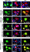
Full-length HPK1 and the isolated HPK1 C terminus localize comparably in Cos1 cells. (Top left) Schematic representation of the domain structure of HPK1-HA and the HPK1 C terminus fused to EGFP (EGFP-HPK1291-827). In EGFP-HPK1291-827, the kinase domain (kinase dom.) has been replaced by EGFP. P1, P2, and P4 denote proline-rich SH3 domain-binding sites. Tyrosine 379 can be phosphorylated by Src/Syk family kinases and provides a docking site for the SH2 domain of SLP-76 family adaptors. The numbers are amino acid positions in HPK1. The epitope tag (HA) is indicated. HPK1-HA (a to f), EGFP (g and h), or the EGFP-HPK1291-827 hybrid protein (i to o) was transiently expressed in Cos1 cells grown on coverslips. For visualization of HPK1-HA, formaldehyde-fixed cells were stained with anti-HA antibodies and then with Alexa Fluor 488-labeled secondary antibodies. Cells were embedded in fluorescence mounting medium (Mowiol) and observed with an epifluorescence microscope using a 63×/1.25 oil immersion objective (b, c, and e to o; bars, 25 μm) or a 20× objective (a and d; bars, 100 μm). HPK1-HA (a to c) and EGFP-HPK1291-827 (g, i, k, and l) are either shown alone or as an overlay with nuclei counterstained by Hoechst 33258 (d, e, f, h, m, n, and o). (a to c) Cells expressing low (L), medium (M), or high (H) levels of HPK1-HA (arrowheads) are identified. Cells expressing low, medium, or high levels of EGFP-HPK1291-827 are shown in panel i, k, or l, respectively.
FIG. 2.
FIG. 2.
The adaptor protein Clnk enhances phosphorylation of HPK1 on tyrosine in the presence of the active Src kinase Lck. (A) The EGFP-HPK1291-827 fusion protein is not subject to excessive proteolysis in Cos1 cells. Expression plasmids for EGFP, EGFP-HPK1291-827, or the HPK1 C-terminus HPK1291-827 were transfected into Cos1 cells as indicated. After 24 h, the cells were lysed, and the stability of the HPK1 hybrid protein was tested by anti-HPK1 or anti-EGFP Western blotting. An extraordinarily strong exposure of the EGFP Western blot is shown to visualize degradation products of the HPK1 hybrid protein. (B) Cos1 cells were transfected with expression plasmids encoding either wild-type HPK1 [HPK1(wt)], HPK1(wt) plus Lck, or HPK1(wt) plus Lck and Clnk. After immunoprecipitation, HPK1 tyrosine phosphorylation was assessed by antiphosphotyrosine (anti-pTyr) Western blotting. In parallel, HPK1 kinase activity was determined by an in vitro kinase assay. HPK1 expression was demonstrated by anti-HPK1 Western blotting.
FIG. 3.
FIG. 3.
Active Lck and Clnk direct HPK1-HA or the EGFP-HPK1291-827 fusion protein to the plasma membrane. Cos1 cells grown on coverslips were transfected with expression plasmids encoding either HPK1-HA, Clnk, or Lck alone (top row) or HPK1-HA in combination with Lck or Clnk or Lck and Clnk or EGFP-HPK1291-827 in combination with Lck and Clnk. Twenty-four hours after transfection, cells were fixed with formaldehyde and stained with the respective antibodies. Hoechst 33258 was used to counterstain nuclei (middle column and top row). Specimens were observed with an epifluorescence microscope using a 63×/1.25 oil immersion objective, and micrographs were acquired using a Spot digital camera. Merging of green (anti-HA or anti-EGFP) and red (anti-Clnk or anti-Lck) fluorescence is depicted as overlay.
FIG. 4.
FIG. 4.
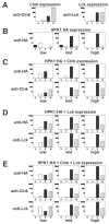
Active Lck dominates HPK1 localization upon overexpression in Cos1 cells. Cos1 cells grown on coverslips were transfected with expression plasmids encoding either Clnk or Lck (A) or HPK1-HA (B), HPK1-HA plus Clnk (C), HPK1-HA plus Lck (D), or HPK1-HA plus Clnk and Lck (E) as described in the legend to Fig. 3. Twenty-four hours after transfection, cells were fixed and stained with antibodies directed against the HA tag, Clnk, or Lck. In each experimental group, successfully transfected cells were then classified as expressing low, middle, or high levels of HPK1 (Fig. 1). For at least 20 cells of each level of expression of HPK1, HPK1 fluorescence in the nucleus (N), cytoplasm (C), and spots in the cytoplasm (S) and at the plasma membrane (M) was graded. The level of fluorescence is indicated on the y axes as follows: weak (•), intermediate (••), and strong (•••). The average fluorescence intensity of the different cellular compartments was then blotted for cells expressing low, middle, or high levels of HPK1. When Clnk and/or Lck were coexpressed, intracellular distribution of these proteins was determined in an analogous manner; however, the levels of expression always refer to HPK1 expression.
FIG. 5.
FIG. 5.
Intracellular localization of HPK1 is mediated exclusively by its C-terminal part in Cos1 cells. Cos1 cells grown on coverslips were transfected with expression plasmids encoding either EGFP (A) or EGFP-HPK1291-827 (B) alone or EGFP-HPK1291-827 with Clnk (C), Lck (D), or Clnk and Lck (E) as described in the legend to Fig. 3. Twenty-four hours after transfection, cells were fixed and stained with antibodies directed against Clnk or Lck. In each experimental group, successfully transfected cells were then classified as expressing low, middle, or high levels of EGFP or EGFP-HPK1291-827 as judged by their green fluorescence (Fig. 1). For at least 20 cells of each expression level, green fluorescence (GF) in the nucleus (N), cytoplasm (C), and spots in the cytoplasm (S) and at the plasma membrane (M) was graded. The level of fluorescence is indicated on the y axes as follows: weak (•), intermediate (••), and strong (•••). The average fluorescence intensity of the different cellular compartments was thenblotted for cells expressing low, middle, or high levels of HPK1. When Clnk and/or Lck were coexpressed, intracellular distribution of these proteins was determined in an analogous manner; however, levels of expression always refer to EGFP or EGFP-HPK1291-827 expression.
FIG. 6.
FIG. 6.
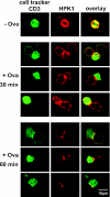
HPK1 is recruited to the contact site of APC-T-cell conjugates. A20 B cells labeled with cell tracker green were pulsed with OVA peptide and incubated with antigen-specific DO11.10 T hybridoma cells to induce conjugate formation. Conjugates were fixed and stained for HPK1 (red) and CD3 (green). In areas where HPK1 is recruited to the contact sites, the overlay of Cy3-stained HPK1 and FITC-stained CD3 and/or cell tracker appears yellow. The negative control shows A20 cells not pulsed with OVA peptide (− Ova); conjugate formation with OVA-pulsed A20 cells was allowed to proceed for 30 or 60 min. Representative conjugates analyzed by confocal laser scanning microscopy are shown.
FIG. 7.
FIG. 7.
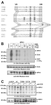
Mutation of potential phosphorylation sites in the HPK1 kinase domain activation loop impairs HPK1 kinase activity and generates HPK1 mutants refractory to TCR ligation-mediated activation. (A) Alignment of the amino acid sequences of activation loops of different serine/threonine kinases. Residues reported to be phosphorylated and/or implicated in the regulation of the respective kinase are shown in bold type. The alignment identified three potential phosphorylation sites in the HPK1 activation loop, which are shown as white letters on a black background. The signature motifs of the kinase subdomains VII (DFG) and VIII (APE) and a central serine/threonine and an invariant threonine residue are highlighted by light grey shading. (B) Cos1 cells were transiently transfected with plasmids encoding Clnk and either empty vector (control) or HPK1 [wild-type (wt)] or the ATP-binding site mutant HPK1(K46E) or various HPK1 constructs containing single or multiple activation loop mutations at position 165 (threonine to alanine [T165A]), position 171 (serine to alanine [S171A]), or position 175 (threonine to alanine [T175A]). After immunoprecipitation, the indicated variant proteins were tested for their abilities to autophosphorylate and to transphosphorylate a recombinant GST-c-JunN fusion protein in an in vitro kinase assay. Reaction products were separated by SDS-PAGE, and phosphoproteins were visualized by autoradiography (top panel). The migratory positions of HPK1 proteins, coimmunoprecipitated Clnk, and GST-c-JunN are indicated to the right of the gels. The levels of expression of HPK1 and Clnk were visualized by Western blotting (bottom panels). A representative experiment of four experiments is shown. (C) Jurkat T cells were transiently cotransfected with plasmids encoding Clnk and either empty vector (control), or HPK1 (wild type [wt]), or HPK1 constructs containing the indicated activation loop mutations. Forty-eight hours after transfection, cells were stimulated for 3 min using the anti-CD3 antibody OKT3. HPK1 proteins were immunoprecipitated and tested for their ability to autophosphorylate in an in vitro kinase assay. Reaction products were separated by SDS-PAGE; the migratory positions of HPK1 proteins and coimmunoprecipitating Clnk are indicated to the right of the gels. Expression of the HPK1 proteins was determined by anti-HA Western blotting. The positions of the two nonspecific signals are indicated by asterisks. The experiment was repeated twice with similar results.
FIG. 8.
FIG. 8.
HPK1 activation loop mutants are severely impaired in mediating SAPK/JNK and NF-κB activation. (A) Wild-type HPK1 (wt) or HPK1 proteins bearing the indicated activation loop mutations were transiently expressed in Cos1 cells together with p54-SAPKβ/JNK. Forty-eight hours after transfection, coexpressed SAPK/JNK protein was immunopurified, and then its kinase activity towards bacterially expressed GST-c-JunN protein was determined. Phosphorylated reaction products were separated by SDS-PAGE, and relative GST-c-JunN phosphorylation levels were determined assessed using a phosphorimager. Expression of HPK1 proteins was demonstrated by anti-HPK1 Western blotting. A representative experiment of two experiments is shown. (B) Cos1 cells were transfected with increasing amounts of plasmids (1, 2, and 4 μg) encoding either wild-type HPK1 (wt) or HPK1 constructs containing single activation loop mutations as indicated or empty vector (−) in the presence of a NF-κB double reporter system (2). After 48 h, NF-κB-driven luciferase activity was determined and normalized against a β-galactosidase transfection control using a chemiluminescence assay system. Relative activation of NF-κB-driven luciferase activity normalized for transfection efficacy is shown. Bars depict the averages of duplicate samples; the experiment was repeated three times with similar results. Matching levels of expression of HPK1 proteins were visualized by anti-HPK1 Western blotting.
FIG. 9.
FIG. 9.
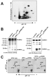
HPK1 proteins autophosphorylate in vitro on multiple amino acids. Autophosphorylation sites in the kinase domain are mostly threonine residues. (A) HPK1 protein was transiently expressed in Cos1 cells and subjected to immunoprecipitation and autophosphorylation in vitro. Reaction products were separated by SDS-PAGE and visualized by autoradiography. Full-length HPK1 phosphoprotein was eluted from the gel (white box in panel B) and subjected to tryptic digestion. Tryptic phosphopeptides were separated by two-dimensional TLC on a Hunter HLTE-7000 machine and visualized by autoradiography. The polarity of the electrophoretic separation and direction of the ascending chromatography are indicated. (B) Full-length HA-tagged HPK1 (wild type) was coexpressed in Cos1 cells with either empty vector (control) or with the HA-tagged HPK11-232 truncation mutant consisting of the first 232 amino acids of HPK1. The truncation mutant was either wild type at amino acid position 46 or harbored a K46E mutation which precludes ATP binding to the kinase domain. Anti-HA immunoprecipitation and a subsequent in vitro kinase reaction demonstrated transphosphorylation of the kinase-dead HPK11-232 fragment (left panel). Expression of all HPK1 proteins was visualized by anti-HA Western blotting (right panel). The white box depicts full-length HPK1 protein eluted for the generation of tryptic phosphopeptide maps as shown in panel A. The black box depicts the HPK11-232 fragment that was eluted for the subsequent phosphoamino acid analysis (C) and tryptic phosphopeptide maps (Fig. 10B). (C) HPK1 (wild type [wt]) or truncated HPK11-232(K46E) or HPK11-232(K46E) containing a triple activation loop mutation (TST165,171,175AAA) were transphosphorylated by full-length HPK1 in vitro as described in panel B. After SDS-PAGE, the depicted HPK1 phosphoproteins were eluted and subjected to phosphoamino acid analysis using a Hunter HTLE-7000 machine. The buffer system, polarity of the electrophoretic separations, and positions of reference phosphoamino acids are indicated. p-Ser, phosphoserine; p-Thr, phosphothreonine; p-Tyr, phosphotyrosine, Pi, free phosphate.
FIG. 10.
FIG. 10.
Phosphotryptic peptide mapping identifies threonine 165 in the HPK1 activation loop as an autophosphorylation site. (A) Wild-type HPK11-232(K46E)-HA (wt) or HPK11-232(K46E)-HA proteins containing the indicated activation loop mutations were transiently coexpressed with full-length kinase-competent HPK1-HA. After immunopurification using anti-HA antibodies, the truncated proteins were transphosphorylated by full-length HPK1 in vitro. Reaction products were separated by SDS-PAGE, phosphoproteins were visualized by autoradiography, and expression of the different truncated proteins was demonstrated by anti-HA Western blotting. The various mutations in the activation loop sequence are indicated over the gel. (B) The HPK11-232-HA proteins shown in panel A were recovered from the SDS-polyacrylamide gel and subjected to tryptic peptide mapping. Phosphopeptides were separated on a Hunter HTLE-7000 machine and visualized by autoradiography. The maps are oriented as in Fig. 9A. (C) HPK1 activation loop amino acid sequence. The potentially phosphorylated amino acids threonine 165, serine 171, and threonine 175 (asterisks) and trypsin cleavage sites (scissors) are indicated. The sequences of four possible tryptic peptides generated from this sequence are displayed. The charges of charged amino acids at pH 1.9 are denoted. (D) Phosphoamino acid analysis of the phosphopeptides 1 and 2 indicated in panel b of panel B. The spots corresponding to peptides 1 and 2 were identified by autoradiography and scraped off the TLC plate, and phosphopeptides were eluted. After digestion in 6 M HCl, phosphoamino acids were separated. The buffer system, polarity of the electrophoretic separations, and positions of reference phosphoamino acids are indicated as described in the legend to Fig. 9C.
FIG. 11.
FIG. 11.
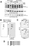
Serine 171 in the HPK1 activation loop can be transphosphorylated by PKD1. (A) The indicated HPK11-232(K46E)-HA truncation proteins were transiently coexpressed with activated versions of full-length PKD1-HA. Anti-HA immunopurified proteins were subjected to in vitro kinase reactions and separated by SDS-PAGE. The migratory position of the HPK11-232 fragment is indicated to the right of the gels. Expression of the different proteins was verified by Western blotting. wt, wild-type protein. (B) (Left) HPK11-232(K46E)-HA protein was transphosphorylated by the PKD1 (Zn-Mut) mutant in vitro. After separation of the reaction mixture by SDS-PAGE, the HPK1 fragments were recovered from the SDS-polyacrylamide gel and subjected to tryptic peptide mapping. Phosphopeptides were separated on a Hunter HTLE-7000 machine and visualized by autoradiography. The maps are oriented as in Fig. 9A. (Right) Schematic depiction of the phosphotryptic map on the left. Major spots which coincide with the presence of S171 in the activation loop peptide are shown in black; these spots are not present after phosphorylation of the HPK11-232(K46E,S171T)-HA fragment. Dark grey spots likely represent degradation products of the active kinase PKD1 (Zn-Mut). (C) Phosphoamino acid analysis of phosphopeptides 3 and 4 indicated in the right panel of panel B. The buffer system, polarity of the electrophoretic separations, and positions of reference phosphoamino acids are indicated as described in the legend to Fig. 9C. (D) Activation loop sequence alignment of subfamily I GCKs. Grey boxes highlight the amino acids T165 and S171 that are phosphorylated in and contribute to the activation of HPK1. A hydrophobic residue at position −5 relative to S171 (white letters on black background) is critical for phosphorylation by PKD1 and conserved in all family members.
FIG. 12.
FIG. 12.
PKD influences HPK1 kinase activity in various cell types and augments HPK1 effector pathway signal strength. (A) HPK1-HA and wild-type PKD1 [PDK1(WT)], the dominant-active PKD1 variants PKD1 (PH-Mut) or PKD1 (Zn-Mut) or the kinase-dead mutant PKD1 (KD) were transiently expressed in Jurkat T cells. After 48 h, samples were split, and half of the cells were stimulated for 3 min by anti-CD3 (αCD3) TCR ligation. Cells were lysed, and HPK1 protein was immunopurified and subjected to an in vitro kinase reaction. HPK1 activity was determined in the presence of a recombinant GST-c-JunN fusion protein as a pseudosubstrate. The activation values describe the increases in HPK1 kinase activity in the presence (+) or absence (−) of CD3 ligation. The experiment was repeated three times with similar results. (B) 293T cells were transfected with an HPK1-HA expression plasmid and either empty vector or a PKD1 antisense (PKD1 a.s.) plasmid at a 1:1 or 1:2 ratio. The activity of HPK1 protein was determined as described in the legend to Fig. 7B. Immunoprecipitated HPK1-HA protein (IP) was assessed by anti-HA Western blotting. PKD levels of expression and protein content of the lysates were visualized by anti-PKD and antitubulin Western blotting. We show one of three experiments; the three experiments had the same outcome. (C) Mouse DC27.1 T cells were transfected with empty vector or a PKD1 antisense (PKD1 a.s.) plasmid. Forty-eight hours after transfection, cells were stimulated for 3 min with anti-CD3 antibody (+) or left unstimulated (−). Endogenous HPK1 proteins were immunoprecipitated and tested for their ability to autophosphorylate in an in vitro kinase assay. HPK1 protein present in the immunoprecipitation (IP) was controlled by anti-HPK1 Western blotting. The levels of expression in the protein lysate were visualized by anti-PKD1 and antitubulin Western blotting. One of three experiments is shown; the three experiments had similar results. (D) HPK1, wild-type PKD1 [PKD1(WT)], or kinase-dead PKD1 (KD) were transiently expressed in Cos1 cells as indicated. The activity of coexpressed SAPK/JNK protein was determined as described in the legend to Fig. 8A. The levels of expression were visualized by anti-SAPK, anti-HPK1, or anti-PKD1 Western blotting. Bars represent the averages of duplicate transfection experiments. A representative experiment is depicted. The two experiments had similar results. (E) Cos1 cells were transfected with increasing amounts of HPK1 expression plasmid (1, 2, and 4 μg) and either a constant amount of constitutively active PKD1 (PH-Mut) or empty vector in the presence of a 8x NF-κB double reporter system. Analysis was done as described in the legend to Fig. 8B. The levels of expression were visualized by anti-HPK1 or anti-PKD1 Western blotting. The experiment was repeated twice with identical results.

References

    1. Anafi, M., F. Kiefer, G. D. Gish, G. Mbamalu, N. N. Iscove, and T. Pawson. 1997. SH2/SH3 adaptor proteins can link tyrosine kinases to a Ste20-related protein kinase, HPK1. J. Biol. Chem. 272:27804-27811. - PubMed
    1. Arnold, R., J. Liou, H. C. Drexler, A. Weiss, and F. Kiefer. 2001. Caspase-mediated cleavage of hematopoietic progenitor kinase 1 (HPK1) converts an activator of NFκB into an inhibitor of NFκB. J. Biol. Chem. 276:14675-14684. - PubMed
    1. Biondi, R. M. 2004. Phosphoinositide-dependent protein kinase 1, a sensor of protein conformation. Trends Biochem. Sci. 29:136-142. - PubMed
    1. Boyle, W. J., P. van der Geer, and T. Hunter. 1991. Phosphopeptide mapping and phosphoamino acid analysis by two-dimensional separation on thin-layer cellulose plates. Methods Enzymol. 201:110-149. - PubMed
    1. Chadee, D. N., T. Yuasa, and J. M. Kyriakis. 2002. Direct activation of mitogen-activated protein kinase kinase kinase MEKK1 by the Ste20p homologue GCK and the adapter protein TRAF2. Mol. Cell. Biol. 22:737-749. - PMC - PubMed

Publication types

MeSH terms