. 2005 Mar;25(6):2419-30.
doi: 10.1128/MCB.25.6.2419-2430.2005.

p21WAF1/CIP1 selectively controls the transcriptional activity of estrogen receptor alpha

Affiliations

p21WAF1/CIP1 selectively controls the transcriptional activity of estrogen receptor alpha

Asmaà Fritah et al. Mol Cell Biol. 2005 Mar.

Abstract

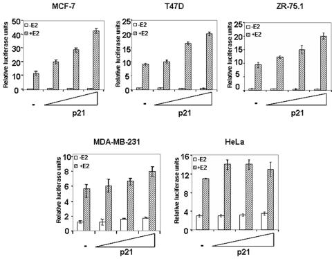
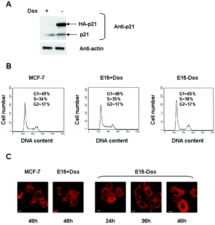
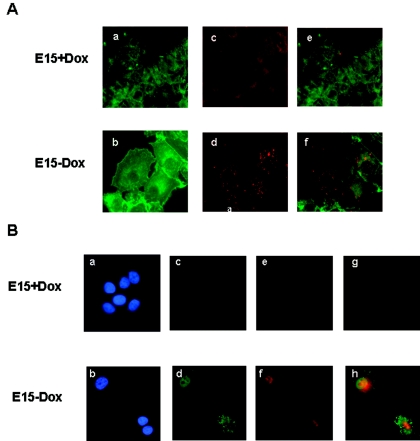
Estrogen receptors (ER) are ligand-dependent transcription factors that regulate growth, differentiation, and maintenance of cellular functions in a wide variety of tissues. We report here that p21WAF1/CIP1, a cyclin-dependent kinase (Cdk) inhibitor, cooperates with CBP to regulate the ERalpha-mediated transcription of endogenous target genes in a promoter-specific manner. The estrogen-induced expression of the progesterone receptor and WISP-2 mRNA transcripts in MCF-7 cells was enhanced by p21WAF1/CIP1, whereas that of the cyclin D1 mRNA was reduced and the pS2 mRNA was not affected. Chromatin immunoprecipitation assays revealed that p21WAF1/CIP1 was recruited simultaneously with ERalpha and CBP to the endogenous progesterone receptor gene promoter in an estrogen-dependent manner. Experiments in which the p21WAF1/CIP1 protein was knocked down by RNA interference showed that the induction of the expression of the gene encoding the progesterone receptor required p21WAF1/CIP1, in contrast with that of the cyclin D1 and pS2 genes. p21WAF1/CIP1 induced not only cell cycle arrest in breast cancer cells but also milk fat globule protein and lipid droplets, indicators of the differentiated phenotype, as well as cell flattening and increase of the volume of the cytoplasm. These results indicate that p21WAF1/CIP1, in addition to its Cdk-regulatory role, behaves as a transcriptional coactivator in a gene-specific manner implicated in cell differentiation.


Figures

FIG. 1.
FIG. 1.
The transcriptional effect of p21 varies according to the cell line. Cells were transfected with increasing amounts (0.5, 1, or 2 μg) of pCMV-p21 along with 0.5 μg of ERE-tk-luc and 0.1 μg of a β-galactosidase construct as an internal control. In the ERα-negative cells, MDA-MB-231 and HeLa cells, the transfection mix included 0.1 μg of pSG5-hERα. After overnight incubation, cells were treated with 10 nM estradiol (+E2) or vehicle (−E2) and harvested 24 h later. Extracts were assayed for luciferase and β-galactosidase activities. Luciferase activity was normalized to β-galactosidase activity. The results are shown as means ± standard errors from at least three separate experiments.
FIG. 2.
FIG. 2.
ERα interacts with p21 and CBP. (A) MCF-7 cells were transfected with increasing amounts (0.5, 1, or 2 μg) of pCMV-p21 or pCMV-p27, 1 μg of pRSV-CBP alone or in combination with 1 μg of pCMV-p21, or 1 μg of pCMV-p27 along with 0.5 μg of ERE-tk-luc and 0.1 μg of a β-galactosidase construct as an internal control. (B) MCF-7 cells were electroporated with 20 μg of pCMV-p21 or TranSilent human CBP-siRNA vector alone or in combination with 2 μg of ERE-tk-luc. Total lysates of the control or siRNA-transfected cells were analyzed by SDS-PAGE and immunoblotting for CBP and β-actin. For panels A and B, after overnight incubation, cells were treated with 10 nM estradiol (+E2) or vehicle (−E2) and harvested 24 h later. Extracts were assayed for luciferase and β-galactosidase activities. Luciferase activity was normalized to β-galactosidase activity except for panel B, where all luciferase values were normalized to protein concentration. The data are means ± ranges of duplicates and illustrate one of three experiments which all gave similar results. (C) Equal amounts of 35S-labeled p21 or p27 were incubated with bacterially expressed GST or GST-ERα fusion constructs immobilized on glutathione-Sepharose beads. Following incubation at 4°C for 2 h, complexes were washed five times with binding buffer, resolved by SDS-PAGE, and detected by fluorography. As a control, 10% of the 35S-labeled protein used in the experiment was analyzed in parallel (Input). (D) MCF-7 cells were maintained in phenol red-free medium supplemented with 10% dextran-charcoal-stripped serum for 48 h and then grown in medium with or without 10 nM estradiol for 2 h. For immunoprecipitations, 500 μg of nuclear extract was incubated with the anti-CBP antibody or control normal rabbit IgG (lanes 3 to 5). After incubation with 10 mM dithiothreitol at 37°C for 30 min, the supernatant was collected and reimmunoprecipitated with antibodies against p21 or control normal mouse IgG (lanes 6 to 8). The immunoprecipitates were subjected to SDS-PAGE and immunoblotting. A portion of the nuclear extracts was also analyzed by Western blotting with antibodies against ERα, CBP, and p21 (Input) (lanes 1, 2).
FIG. 3.
FIG. 3.
p21 selectively modulates estrogen-dependent transcription. (A) MCF-7 cells electroporated with empty vector or p21 expression vector were incubated in hormone-free medium for 48 h and then stimulated with 10 nM estradiol for 1, 4, or 8 h or left untreated. mRNA levels were analyzed by real-time RT-QPCR, as described in Materials and Methods, with primers specific for ERα target genes. The results after standardization to 36B4 in the same experiments are shown and are the means ± standard errors of triplicates. (B) MCF-7 cells were electroporated as for panel A and placed in medium containing or lacking 10 nM estradiol for 24 h. Cell extracts were prepared and tested for PRB, PRA, pS2, cyclin D1, p21, and ERα proteins by immunoblotting.
FIG. 4.
FIG. 4.
Endogenous p21 is required for ERα function. (A) MCF-7 cells were electroporated with 20 μg of p21-siRNA400, p21-siRNA225, or scrambled siRNA together with 2 μg of ERE-tk-luc and 0.8 μg of a β-galactosidase construct as an internal control. After overnight incubation, cells were treated with 10 nM estradiol (+E2) or vehicle (−E2) and harvested 24 h later. Extracts were assayed for luciferase and β-galactosidase activities. Luciferase activity was normalized to β-galactosidase activity. Values shown are means ± standard errors from at least three separate experiments. Total lysates of the siRNA-transfected cells were analyzed by SDS-PAGE and immunoblotting for p21 and β-actin. (B) MCF-7 cells were electroporated with 20 μg of p21-siRNA400 or scrambled-siRNA vectors. Forty-eight hours after transfection, cells were treated with 10 nM estradiol for 8 h or left untreated. mRNAs were analyzed by real-time RT-QPCR to measure the levels of p21, PRB, pS2, and cyclin D1 transcripts. C. MCF-7 cells were electroporated and treated as in panel B. Cell extracts were prepared and tested for ERα, PRB, pS2, and cyclin D1 proteins by immunoblotting.
FIG. 5.
FIG. 5.
p21 is recruited to the PR promoter in an estrogen-dependent manner. (A) Cross-linked, sheared chromatin from MCF-7 cells incubated with or without estradiol (45 min) was immunoprecipitated with the indicated antibodies. DNA was analyzed by PCR, using primers to amplify the pS2 promoter, the cyclin D1 promoter, or two different regions of the PR promoter. Results shown are representative of four independent experiments. (B) Real-time quantitative PCR analysis. Results are shown as percentages of input (means ± standard errors of triplicates). PRB, PR promoter region −125 to +187; PRA, PR promoter region +352 to +616.
FIG. 6.
FIG. 6.
p21 inhibits proliferation and induces morphological changes in E15 cells. (A) Induction of exogenous p21 by removal of DOX in the E15 clone. The cells were cultured for 48 h in the presence or absence of DOX and harvested for analysis of HA-p21 expression by Western blotting with an anti-p21 antibody. β-Actin was used as a control. (B) E15 cells were cultured in the presence or absence of DOX for 48 h. Cells were harvested, fixed with ethanol, and stained with propidium iodide. Cell cycle distribution was then determined by flow cytometry. Cell number is plotted against DNA content. (C) MCF-7 cells and E15 cells were cultured with or without DOX for the indicated times. Cytokeratin 18 detection using a Texas Red-tagged secondary antibody is shown. Images were obtained by microscopy (magnification, ×63). Scale bars, 7 μm. The results were verified in three experiments.
FIG. 7.
FIG. 7.
p21 induces lipid accumulation and milk fat proteins in E15 cells. (A) E15 cells were cultured with (a, c, and e) or without (b, d, and f) DOX for 48 h. Cells were fixed, permeabilized, and then sequentially incubated with fluorescein-phalloidin to identify actin filaments (a and b) and Nile red to identify lipid droplets (c and d). Cells which fluoresce both green and red appear orange (e and f). (B) E15 cells were cultured with (a, c, e, and g) or without (b, d, f, and h) DOX for 48 h. Cells were fixed, permeabilized, and then incubated with either anti-HA or anti-MFG antibody. HA-p21 was detected using a fluorescein-labeled secondary antibody (c and d), and MFGs were detected using a Texas Red-labeled secondary antibody (e and f). Nuclear DNA was stained with DAPI (a and b). Cells which fluoresce both green and red appear orange (g and h). Images were obtained by microscopy (magnification, ×63). The results were confirmed by three independent experiments.
FIG. 8.
FIG. 8.
Proposed model for p21-mediated ERα transactivation. First, p21 inhibits the phosphorylation of CBP by Cdk and thus enhances CBP HAT activity. Second, in the presence of estradiol, p21 binds to CBP and ER on ERE-driven promoters of genes expressed in differentiated cells and favors their transcription.

Similar articles

Cited by

References

    1. Acevedo, M. L., and W. L. Kraus. 2003. Mediator and p300/CBP-steroid receptor coactivator complexes have distinct roles, but function synergistically, during estrogen receptor alpha-dependent transcription with chromatin templates. Mol. Cell. Biol. 23:335-348. - PMC - PubMed
    1. Albanese, C., M. D'Amico, A. T. Reutens, M. Fu, G. Watanabe, R. J. Lee, R. N. Kitsis, B. Henglein, M. Avantaggiati, K. Somasundaram, B. Thimmapaya, and R. G. Pestell. 1999. Activation of the cyclin D1 gene by the E1A-associated protein p300 through AP-1 inhibits cellular apoptosis. J. Biol. Chem. 274:34186-34195. - PubMed
    1. Andrews, N. C., and D. V. Faller. 1991. A rapid micropreparation technique for extraction of DNA-binding proteins from limiting numbers of mammalian cells. Nucleic Acids Res. 19:2499. - PMC - PubMed
    1. Brouillard, F., and C. E. Cremisi. 2003. Concomitant increase of histone acetyltransferase activity and degradation of p300 during retinoic acid-induced differentiation of F9 cells. J. Biol. Chem. 278:39509-39516. - PubMed
    1. Brummelkamp, T. R., R. Bernards, and R. Agami. 2002. A system for stable expression of short interfering RNAs in mammalian cells. Science 296:550-553. - PubMed

Publication types

MeSH terms