. 2005 Mar;25(6):2463-74.
doi: 10.1128/MCB.25.6.2463-2474.2005.

Novel checkpoint response to genotoxic stress mediated by nucleolin-replication protein a complex formation

Affiliations

Novel checkpoint response to genotoxic stress mediated by nucleolin-replication protein a complex formation

Kyung Kim et al. Mol Cell Biol. 2005 Mar.

Abstract

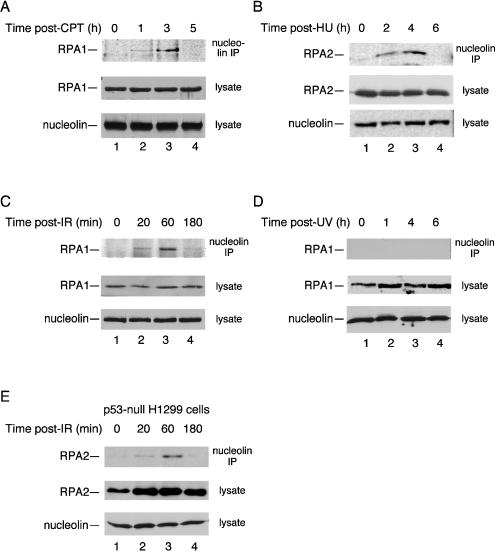
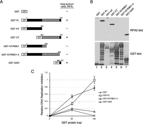
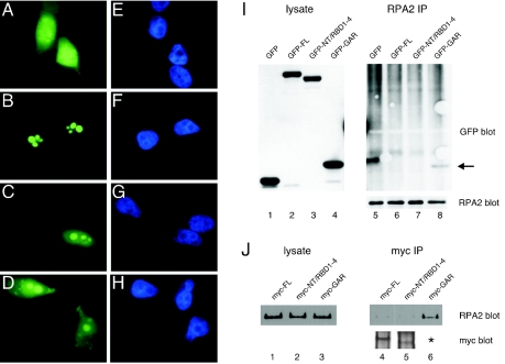
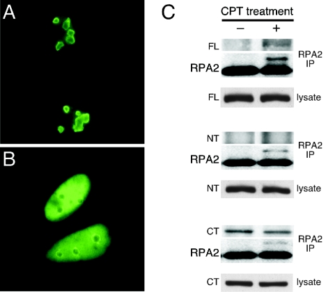
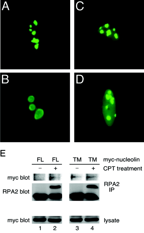
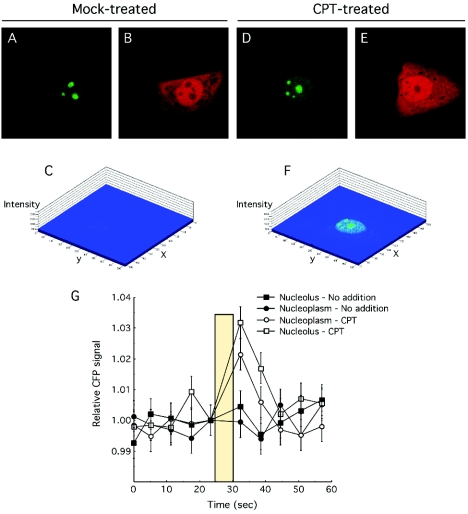
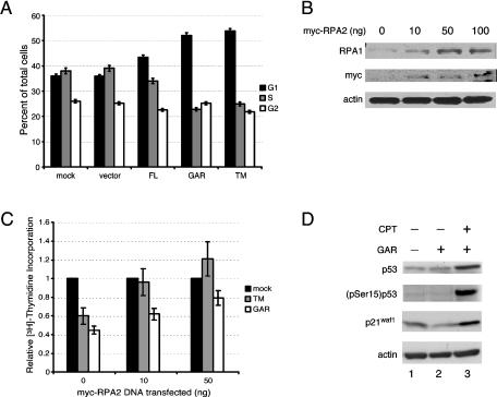
Human replication protein A (RPA), the primary single-stranded DNA-binding protein, was previously found to be inhibited after heat shock by complex formation with nucleolin. Here we show that nucleolin-RPA complex formation is stimulated after genotoxic stresses such as treatment with camptothecin or exposure to ionizing radiation. Complex formation in vitro and in vivo requires a 63-residue glycine-arginine-rich (GAR) domain located at the extreme C terminus of nucleolin, with this domain sufficient to inhibit DNA replication in vitro. Fluorescence resonance energy transfer studies demonstrate that the nucleolin-RPA interaction after stress occurs both in the nucleoplasm and in the nucleolus. Expression of the GAR domain or a nucleolin mutant (TM) with a constitutive interaction with RPA is sufficient to inhibit entry into S phase. Increasing cellular RPA levels by overexpression of the RPA2 subunit minimizes the inhibitory effects of nucleolin GAR or TM expression on chromosomal DNA replication. The arrest is independent of p53 activation by ATM or ATR and does not involve heightened expression of p21. Our data reveal a novel cellular mechanism that represses genomic replication in response to genotoxic stress by inhibition of an essential DNA replication factor.


Figures

FIG. 1.
FIG. 1.
Nucleolin-RPA complex formation is induced after genotoxic stress. Cell lysates were prepared from p53-positive U2-OS cells (A to D) and p53-null H1299 cells (E) at various times after exposure to various stress treatments as follows: 1 μM CPT for 1 h (A), 2.5 mM hydroxyurea (HU) for 1 h (B), 10 Gy of ionizing radiation (IR) (C and E), and UV irradiation with a single dose of 30 J m−2 (D). After each time point, nucleolin was immunoprecipitated from the lysate with a mouse monoclonal antibody to nucleolin. The precipitate was subjected to SDS-PAGE and immunoblotted for RPA with either an anti-RPA1 or anti-RPA2 antibody (as indicated). As loading controls, aliquots of the lysates were subjected to immunoblotting with anti-nucleolin, anti-RPA1, or anti-RPA2 antibodies.
FIG. 2.
FIG. 2.
The nucleolin RPA-binding domain inhibits SV40 DNA replication in vitro. (A) Schematic showing GST-tagged nucleolin and nucleolin mutant proteins as follows: full length (FL), amino terminus (NT), carboxy terminus (CT), amino terminus including the first RBD (NT/RBD1), the GAR deletion mutant (NT/RBD1-4), and only the C-terminal GAR domain (GAR). For each construct, the N-terminal acidic domain is indicated in dark gray; each of the four RBDs have light gray shading and are numbered, and the GAR domain is shown in black. (B) Far-Western analysis of the nucleolin-RPA interaction. Equivalent amounts (500 ng) of nucleolin FL (lane 2), NT (lane 3), CT (lane 4), NT/RBD1 (lane 5), NT/RBD1-4 (lane 6), and GAR (lane 7), with each containing an N-terminal GST tag, were separated by SDS-PAGE. GST alone was also electrophoresed as a control (lane 1). After transfer to a nitrocellulose membrane, the membrane was probed with purified RPA (0.2 μg/ml) (upper panel, lanes 1 to 7). The binding of RPA was visualized by using an RPA2 antibody. To visualize GST-tagged proteins, the membrane was stripped and subjected to immunoblot analysis with a rabbit anti-GST antibody (lower panel, lanes 1 to 7). (C) An SV40 ori-containing plasmid (180 ng) was incubated with AS65 extract (100 μg), T antigen (750 ng), RPA (200 ng), and purified GST-tagged nucleolin proteins (as indicated) for 2 h at 37°C (52). Both FL (□) and GAR (×) GST-tagged nucleolin proteins are proficient in inhibiting SV40 DNA replication in vitro, whereas the NT/RBD1-4 (▵) GST-tagged nucleolin protein and GST alone (⋄) are not. Replication activity was determined by precipitating the reaction mixtures with trichloroacetic acid and determining the amount of 32P in the precipitate by scintillation counting. The data was plotted as the relative DNA replication inhibition compared to that determined by using 100 ng of GST-FL. The maximum degree of inhibition was to 68% that of control levels.
FIG. 3.
FIG. 3.
Complex formation between the nucleolin GAR domain and RPA in vivo. (A to H) U2-OS cells were transfected with GFP alone (A and E) or the GFP-tagged nucleolin derivatives FL (B and F), NT/RBD1-4 (C and G), or GAR (D and H). At 24 h posttransfection, cells were fixed by treatment with 4% (wt/vol) formaldehyde for 30 min at room temperature and then imaged by epifluorescence microscopy. The staining patterns of the various GFP constructs are shown (A to D), as are images of the same cells stained with DAPI (4′,6′-diamidino-2-phenylindole) (E to H). (I) Immunoprecipitation of endogenous RPA protein in U2-OS cells expressing GFP-tagged nucleolin FL (lane 6), NT/RBD1-4 (lane 7), or GAR (lane 8) or GFP alone (lane 5). The coprecipitation of the expressed GFP-tagged proteins with RPA is shown in the GFP blot. The arrow points to the coprecipitation of GFP-tagged GAR (lane 8). Corresponding lysates were assayed for similar levels of protein expression by blotting for GFP (lanes 1 to 4), whereas equivalent immunoprecipitation of RPA was verified by blotting for RPA2 (right side, lower panel). (J) Reverse immunoprecipitation experiment showing the coprecipitation of endogenous RPA in U2-OS cells expressing Myc-tagged nucleolin FL (lane 4), NT/RBD1-4 (lane 5), and GAR (lane 6). Myc-tagged nucleolin proteins were immunoprecipitated, and coprecipitation of RPA was determined by blotting for RPA2 (upper panel). The immunoprecipitated Myc-tagged proteins are also shown (lower panel). The asterisk indicates that the Myc-tagged GAR could not be detected because of its small size (5 kDa), preventing binding to nitrocellulose membrane during the transfer step. However, similar levels of myc staining were observed for the three constructs when transfected cells were examined by immunofluorescence microscopy (data not shown). The lysates were also blotted for RPA2 as a control (lanes 1 to 3).
FIG. 4.
FIG. 4.
Complex formation between nucleolin FL and endogenous RPA stimulated by genotoxic stress. (A and B) The cellular localization of GFP-tagged nucleolin FL expressed in U2-OS cells is shown in the absence of CPT treatment (A) and after treatment with 1 μM CPT for 1 h and a 1-h recovery period (B). (C) Immunoprecipitation of endogenous RPA protein with RPA2 antibody in U2-OS cells expressing GFP-tagged nucleolin FL (top set), nucleolin NT (second set), or nucleolin CT (third set). Coprecipitation of the nucleolin proteins was examined in the absence of CPT treatment (−) and 2 h after treatment with 1 μM CPT for 1 h (+) (upper panels). The same blot was reprobed with RPA2 antibody as a control for RPA immunoprecipitation (middle panels). Lysates were assayed for equivalent expression of GFP fusion proteins by probing with an anti-GFP antibody (lower panels).
FIG. 5.
FIG. 5.
The nucleolin TM mutant constitutively interacts with endogenous RPA. The subcellular localization of nucleolin FL (A and B) and nucleolin TM (C and D) in U2-OS cells was determined. At 24 h posttransfection, cells were either mock treated (A and C) or examined 2 h after treatment with 1 μM CPT for 1 h (B and D). Cells were prepared for epifluorescence microscopy as described in Materials and Methods. (E) The coprecipitation of Myc-tagged nucleolin FL and TM with endogenous RPA in U2-OS cells was examined 24 h posttransfection either with or without prior CPT treatment (as described above). Endogenous RPA was precipitated with anti-RPA2 antibody, and the coprecipitation of Myc-tagged nucleolin FL or TM was visualized by Western blotting with an anti-Myc antibody (9E10).
FIG. 6.
FIG. 6.
Nucleolin FL-RPA complex formation occurs both in the nucleolus and in the nucleoplasm after stress. (A to F) U2-OS cells were transfected with CFP-nucleolin and YFP-RPA2 and either mock treated or treated with 1 μM CPT for 1.5 h prior to imaging. Cells were then imaged to capture the CFP-nucleolin signal (A and D), the YFP-RPA2 signal (B and E), or the FRET signal obtained by transfer of the CFP emission energy to YFP (C and F). The FRET images are shown with a pseudo three-dimensional display with the intensity of staining given on the z axis. (G) Acceptor photobleaching analysis of nucleolin-RPA complex formation. The CFP signal from various (ca. 10 to 15) ROIs was determined at 6-s intervals. After the fifth scan, the YFP fluor was photobleached at 514 nm with an average bleach time of 5 s. An increase in the CFP after photobleaching of the YFP signal is indicative of bona fide FRET (33).
FIG. 7.
FIG. 7.
RPA overexpression rescues the inhibition of DNA replication caused by nucleolin GAR or TM. (A) Cell cycle distribution of U2-OS cells transfected with nucleolin and nucleolin mutants. FACS analysis was performed 24 h after transfection with N-terminal Myc-tagged nucleolin constructs. The DNA content was quantitated by using the DNA intercalating agent 7-aminoactinomycin D, and cells in S phase were identified by determining BrdU incorporation. (B) Overexpression of myc-tagged RPA2 leads to a corresponding increase in the level of RPA1. Lysates from U2-OS cells transfected with various amounts of myc-tagged RPA2 (10, 50, and 100 ng) were analyzed by Western blotting for the level of RPA1 (first panel). Myc-tagged RPA2 expression (second panel) and β-actin (third panel) are shown as transfection and loading controls, respectively. (C) [3H]thymidine incorporation assay shows that expression of Myc-tagged RPA2 (leading to higher levels of RPA) can rescue the reduction in DNA synthesis caused by expression of nucleolin TM or nucleolin GAR. Each set of U2-OS cells (mock transfected, nucleolin TM transfected, and nucleolin GAR transfected) were cotransfected with 0, 10, or 50 ng of Myc-tagged RPA2. The data are plotted showing the relative amounts of [3H]thymidine incorporation compared to the mock-treated cells at each level of Myc-RPA2 transfected. Expression of Myc-RPA2 alone slightly inhibited cellular DNA synthesis with transfection of 10 ng of the expression construct causing a 16% reduction in [3H]thymidine incorporation. This resulted from inhibitory effects of pEF6/Myc vector transfection rather than expression of Myc-RPA2 per se (data not shown). (D) The nucleolin-mediated checkpoint response does not involve p53 activation. Lysates from U2-OS cells expressing GFP-tagged nucleolin GAR (lanes 2 and 3) or GFP alone (lane 1) were examined after mock treatment (lanes 1 and 2) or 2 h posttreatment with 1 μM CPT for 1 h (lane 3). Lysates were analyzed by Western blotting for total p53 levels (top panel), p53 phosphorylation at serine 15 (second panel), p21waf1, and the loading control, β-actin.

Similar articles

Cited by

References

    1. Araujo, S. J., and R. D. Wood. 1999. Protein complexes in nucleotide excision repair. Mutat. Res. 435:23-33. - PubMed
    1. Bembenek, J., and H. Yu. 2003. Regulation of CDC14: pathways and checkpoints of mitotic exit. Front. Biosci. 8:d1275-d1287. - PubMed
    1. Biamonti, G., and S. Riva. 1994. New insights into the auxiliary domains of eukaryotic RNA binding proteins. FEBS Lett. 340:1-8. - PubMed
    1. Bivona, T. G., I. Perez De Castro, I. M. Ahearn, T. M. Grana, V. K. Chiu, P. J. Lockyer, P. J. Cullen, A. Pellicer, A. D. Cox, and M. R. Philips. 2003. Phospholipase Cγ activates Ras on the Golgi apparatus by means of RasGRP1. Nature 424:694-698. - PubMed
    1. Block, W. D., Y. Yu, and S. P. Lees-Miller. 2004. Phosphatidyl inositol 3-kinase-like serine/threonine protein kinases (PIKKs) are required for DNA damage-induced phosphorylation of the 32-kDa subunit of replication protein A at threonine 21. Nucleic Acids Res. 32:997-1005. - PMC - PubMed

Publication types

MeSH terms